Download
1 / 53
530 likes | 1.21k Vues
Dissection of the Anterior Compartment of the Arm.

E N D
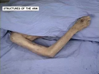
Open the anterior of flexor compartment of the arm by incising the deep fascia in line with the previous skin incisions. Reflect the fascia laterally and medially. Expose the long and short heads of the biceps brachii, coracobrachialis, and brachialis muscles.
Identify the brachial artery; basilic vein;musculocutaneous, median, and ulnar nerves; and the medial cutaneous nerve of the forarm.
Comfirm that the biceps brachii muscle arises by two heads. Note that the long head appears to be shorter than the short head, since it arises from the supraglenoid tubercle of the scapula within the shoulder joint and emerges from the joint beneath the transverse humeral ligment. The short head arises from the tip of the coracoid process of the scapula. Trace the muscle belly down to its tendon in the cubital fossa.
Clean the coracobrachialis muscle from its origin on the tip of the coracoid process of the scapula to its insertion into the medial side of the shaft of the humerus. Note that the muscle is supplied and later pierced by the musculocutaneous nerve.
Clean the brachialis muscle and study its origin from the anterior surface of the shaft of the humerus. It is inserted into the coranoid process of the ulna in the cubital fossa.
Brachial Artery. This is a continuation of the axillary artery at lower border of the teres major muscle. In the cubital fossa it bifurcates into the ulnar and radial arteries.
Clean the brachial artery and note that it is crossed about halfway down the arm by the median nerve and is overlapped on the lateral side by the biceps brachii muscle. It may be easily palpated on the medial side of this muscle in the living. Identify the following branches
1. Muscular branches to the anterior compartment of the upper arm.
2. Profunda brachii artery, which arises near the beginning of the brachial artery and runs with the radial nerve into the posterior compartment of the arm.
3. Superior ulnar collateral artery, which arises near the middle of the upper arm and follows the ulnar nerve.
4. Inferior ulnar collateral artery, which arises near the termination of the artery and takes part in the anastomosis around the elbow joint.
5. Nutrient artery to the humerus, which enters the shaft of the humerus near the insertion of the coracobrachialis muscle.
Basilic Vein. Trace the basilic vein from the front of the elbow to the lower border of the teres major muscle. Here, it joints with the venae comitantes of the brachial artery to form the axillary vein.
Musculocutaneous nerve. Trace this nerve from its origin from the lateral cord of the brachial plexus downward through the coracobrachialis muscle. Follow it between the biceps and brachialis muscle and note that it pierces the deep fascia of the arm just above the elbow at the lateral margin of the biceps tendon. It now continues down the forearm as the lateral cutaneous nerve of the forearm. Identify the branches to the coracobrachialis, biceps, and brachialis muscle.
Median nerve.Trace the median nerve from its origin in the axilla from the medial and lateral cords of the brachial plexus downward into the cubital fossa. Note that it first runs on the lateral side of the brachial artery; then, about halfway down the arm crosses the artery and continues downward on the medial side of the brachial artery. The median nerve has no branches in the upper arm.
Ulnar nerve. Trace the ulnar nerve from the medial cord of the brachial plexus downward on the medial side of the brachial artery as far as the middle of the arm. Note that here, at the insertion of the coracobrachialis, the nerve pierces the medial fascial septum and enters the posterior compartment of the arm. The ulnar nerve has no branches in the anterior compartment of the upper arm.
Medial cutaneous nerve of the forearm. This nerve should be followed from the medial cord of the brachial plexus down the arm on the medial side of the brachial artery. The medial cuatneous nerve of theforearm pierces the deep fascia with the basilic vein and divides into anterior and posterior branches.
Identify the radial nerve as it leaves the axilla and enters the posterior compartment of the arm. Identify it again as it enters the anterior compartment just above the lateral epicondyle of the humerus. Comfirm the existence of the medial and lateral intermuscular fascial septa.
Extend the vertical skin incision on the anterior surface of the arm downward into the cubital fossa. Here, make a transverse incision across the front of the fossa about 5 cm below the level of the medial and lateral epicondyles. Reflect the skin flaps medially and laterally and completely expose the contents of the anterior compartment of the arm and the cubital fossa.
Identify and preserve the cephalic and basilic veins. Study the arrangement of the superficial veins lying anterior to the cubital fossa; they are important. Note that the cubital fossa lies in front of the elbow joint and is triangular in shape.
Confirm that it has the following boundaries:Laterally, the brachioradialis muscle.Medially, the pronator teres muscle.
The base of the cubital fossa triangle is formed by an imaginary line drawn between the two epicondyles of the humerus. The floor of the fossa is formed by the supinator muscle laterally and the brachialis muscle medially. The roof is formed by skin and fascia and is reinforced by the bicipital aponeurosis.
Identify and clean the following structures present within the cubital fossa:
1. The median nerve runs down on the medial side of the brachial artery and leaves the fossa by passing between the two heads of the pronator teres muscle.
2. The brachial artery bifurcates into the radial and ulnar arteries.
3. The tendon of biceps brachii passes to its insertion into the bicipital tuberosity of the radius. Note that the tendon gives off on its medial side the bicipital aponeurosis, which passes downward and medially and fuses with the deep fascia of the forearm.
4. Radial nerve. Trace the radial nerve from the axilla onto the long and medial heads of the triceps and follow it to where it enters the spiral groove on the posterior surface of the humerus. Reidentify the radial nerve as it returns to the anterior compartment of the arm, above the lateral epicondyle of the humerus. Here, it lies between the brachialis and the brachioradialis muscles.
Identify and clean the branches of the radial nerve to the brachialis muscle, brachioradialis muscle, and extensor carpi radialis longus muscle. Identify and clean the two terminal branches of the radial nerve, namely, the deep branch of the radial nerve, which supplies the supinator muscle and then pierces it, and the superficial branch of the radial nerve, which leaves the fossa by passing deep to the branchioradialis muscle.
5. Lateral cutaneous nerve of the forearm.This nerve emerges from beneath the lateral side of the biceps brachii muscle and divides into anterior and posterior branches, which supply the skin of the lateral side of the forearm.
6. Medial cutaneous nerve of the forearm.This nerve runs down the medial side of the brachial artery, pierces the deep fascia about halfway down the arm, and, in the region of cubital fossa, divides into anterior and posterior branches that supply the skin of the medial side of the forearm.
The posterior compartment of the upper arm contains the triceps muscle and its nerve and blood supply. Passing through the compartment are the radial and ulnar nerves.
Make a vertical incision through the deep fascia down the midline of the posterior surface of the arm from the deltoid muscle to the olecranon process of the ulna. By means of transverse incision at the upper and lower ends, reflect medial and lateral flaps to expose the underlying triceps brachii muscle.
Clean the triceps muscle and identify the lateral, long, and medial heads. Follow the muscle to its insertion on the olecranon process of the ulna.
Radial nerve. Expose the radial nerve and the profunda brachii vessels by dividing the lateral head of triceps by means of an oblique incision that runs downward and laterally across the middle of the arm.
Follow the nerve as it lies within the spiral groove and note that it pierces the lateral intermuscular septum to enter the front of the arm.
1. Branches in the axilla. Branches are given to long and medial heads of the triceps, and the posterior cutaneous nerve of the arm is given off.