Download
1 / 42
470 likes | 1.01k Vues
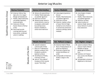
Muscles of the Anterior Forearm. Objectives. Name and identify the muscles in the anterior (flexor/pronator) and posterior (extensor/supinator) compartments of the forearm, noting their relations.

E N D
Objectives • Name and identify the muscles in the anterior (flexor/pronator) and posterior (extensor/supinator) compartments of the forearm, noting their relations. • Name and identify the neurovascular structures in the anterior and posterior compartments of the forearm, noting their relations. • Discuss the functions of the muscles in the anterior and posterior compartment of the forearm.
Functional organization of Anterior and posterior Compartments of forearm.
Names of the Muscles of forearm • Many of them • Long names: good guide to location and function • Location: ie. superficial / deep radial / ulnar (which side is the thumb on??) • Function: ie. flexor / extensor adductor / abductor • Does it act on the thumb (pollux) or the fingers (digits)?
Functional Organization • Anterior compartment – flexor / pronator • Posterior Compartment – extensor / supinator • Flex the wrist ………[flexor] • Extend the wrist ……..[extensor] • Pronate the forearm ……..[pronator] • Acting on wrist (carpus) only ……[carpi] • Acting on fingers ……..[digiti / digitorum] • Acting on thumb (pollux) ……[pollicus]
Flexion / Extension at the wrist joint. Extension Flexion
Adduction / abduction at the wrist joint. Ad-duction Ab-duction
Movements of fingers. MCP (condyloid joints): flexion / extension abduction / adduction IP (hinge joints): flexion / extension X X
Movements of the thumb – a specialized digit, occur at right angles to the fingers. Mainly at the CMC (saddle) joint. MCP – condyloid; IP – hinge (like fingers).
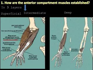
Anterior compartement of forearm Flexor / pronator Compartment • Divided into: • Superficial group(5) • From lateral to medial: • - Pronator teres • Flexor carpi radialis • Palmaris longus • Flexor digitorum • superficialis, • Flexor carpi ulnaris • And :- …… Protonator teres Brachioradialis flexor carpi radialis palmaris longus flexor digitorum superficialis flexor carpi ulnaris
Anterior compartement of forearm Flexor / pronator Compartment • DEEP GROUP (3) • Flexor pollicislongus (FPL) • Flexor digitorumprofundus (FDP) • Pronator quadratus (PQ)
SUPERFICIAL GROUP • ORIGINS: • All 5 muscles have a HUMERAL HEAD (arising from the front of medial epicondyle of humerus: Common flexor origin). • IN ADDITION: • 3 out of 5 muscles have an ULNAR HEAD: • PT & FDS: arise from medial border of coronoid process of ulna • FCU: arises from medial surface of olecranon process & posterior border of shaft of ulna • One muscle has a RADIAL HEAD: • FDS: arises from oblique line & anterior border of shaft of radius
SUPERFICIAL GROUP • Functional organization: • 3 muscles mainly flex at the wrist. • Flexor carpi radialis • Palmaris longus • Flexor carpi ulnaris • Any “carpi” is inserted into metacarpal bone • FCR:(flexion + abduction of wrist joint): inserted into 2nd & 3rd metacarpal bones • FCU:(flexion + adduction of wrist joint): inserted into 5th metacarpal bone (+ pisiform & hamate) • PL:(flexion of wrist joint): inserted into palmar aponeurosis Flexor Carpi Radialis Palmaris Longus Flexor Carpi ulnaris
Pronator Teres • One pronator of radioulnar joints Pronator Teresproduces powerful pronation • Any muscle acting on radioulnar joint must be inserted into radius • Origin: • Medial epicondyle of humerus • Insertion: middle of lateral surface of shaft of radius (point of maximum curvature)
Flexor Carpi Radialis • Origin: • Medial epicondyle of humerus • Insertion: • Base of 2nd & 3rd metacarpals, anterior (palmar surface)
Flexor Carpi Radialis • Action: • Flexion of wrist • Abduction of wrist • Weak flexion of elbow • Innervation: • Median nerve (C6,7)
Palmaris Longus • Origin: • Medial epicondyle of humerus • Insertion: • Palmar aponeurosis of the 2nd, 3rd, 4th, & 5th metacarpals
Palmaris Longus • Action: • Flexion of wrist • Weak flexion of elbow • Innervation: • Median nerve (C6,7)
Flexor DigitorumSuperficialis • Origin: • Humeral head:Medialepicondyle of humerus • Ulnar Head: medial coronoid process • Radial Head: upper 2/3 of anterior border of radius
Flexor DigitorumSuperficialis • Insertion: • by 4 tendons into the middle phalanges of medial 4 fingers • Action: • produces flexion of proximal interphalangeal & metacarpophalangeal joints of medial 4 fingers
Flexor Carpi Ulnaris • Origin: • Medial epicondyle of humerus • Posterior aspect of the proximal ulna • Insertion: • Pisiform, hamate, & base of 5th metacarpal
Flexor Carpi Ulnaris • Action: • Flexion of wrist • Adduction of wrist • Weak flexion of elbow • Innervation: • Ulnar nerve (C8, T1)
DEEP GROUP Flexor PollicisLongus • Origin: • Middle anterior surface of radius(+ interosseous membrane) • Anterior medial border of ulna (just distal to coronoid process) • Insertion: • Base of distal phalanx of thumb (palmar surface)
Flexor PollicisLongus • Action: • Flexion of thumb • Flexion of wrist • Innervation: • Median nerve (C8, T1)
Flexor DigitorumProfundus • Origin: • Proximal ¾ of anterior & medial ulna • Insertion: • Base of distal phalanxes of the 4 fingers
Flexor Digitorum Profundus • Action: • Flexion of fingers • Flexion of wrists • Innervation: Double innervation • Median nerve (C8, T1) to 2nd & 3rd fingers • Ulnar nerve (C8, T1) to 4th & 5th fingers
PRONATOR QUADRATUS • ORIGIN: Lower ¼ of anterior surface of shaft of ulna • INSERTION: Lower ¼ of anterior surface of shaft of radius • ACTION: main pronator of radioulnar joints
Nerve Supply • All forearm muscles are innervated by the MEDIAN nerve EXCEPT: 1 ½ muscles • flexor carpi ulnaris • ulnar side of the flexor digitorumprofundus Plus: All thenarmsexcept adductor pollicis
Anterior Fore Arm Pass Fail Pass Fail
Ant Fore arm Wrist Flexors FCU+FCR FDS FDP PL Wrist Add FCU Wrist Abd FCR Nerve Supply
Post. ForearmSuperficial disection Nerve Supply Radial Nerve Anatomical snuff box
Rotators of the Radius Pronators Supinator Biceps Brachii
Pronation / Supination: Occurs at radio-ulnar jts. Proximal: Head of radius articulates with radial notch of ulna (pivot jt). It is held in place by the annular ligament. Distal – Radius pivots around the fixed distal end of ulna (pivot jt). Radio-ulnar articulation is stabilized by interosseus membrane.
Course of radial and ulnar arteries in forearm Axillary vein: continuation of basilic vein
Radial & Ulnar Arteries medial lateral Radial artery Ulnar artery Common interosseous • Anterior • Posterior Dorsal and palmer carpal branches Dorsal and palmer carpal branches Deep (superficial) palmar arches superficial (deep)palmar arches
RELATION OF ARTERIES, NERVES AND TENDONS AT THE WRIST NANAN Superficial Radial Nerve Radial Artery Median Nerve Ulnar Artery Ulnar Nerve MOVING LATERAL TO MEDIAL 1-9 • Flexor Digitorum Superficialis • Ulnar Artery • Ulnar Nerve • 9. Flexor Carpi Ulnaris • Brachioradialis • Superficial Radial n. • Radial Artery • Flexor Carpi Radialis • 5. Median Nerve Thumb
Neurovasculature(deep). • Lateral: • radial artery • radial nerve • Midline: • median nerve • anterior interosseus a. • anterior interosseus n. • (deep branch of median) • Medial: • ulnar artery • (gives off common interosseus artery divides into anterior and posterior branches) • ulner nerve radial n ulnar a radial a. ulnar n median n anterior interosseus a + n
Radial Nerve Neurovasculature. Radial nerve and its branches supply all muscles in posterior compartment, including brachioradialis (!). - superficial radial nerve - deep radial nerve - posterior interosseus nerve. Posterior interosseus artery runs between superficial and deep muscles Brachioradialis Deep branch posterior interosseus branch Superficial branch
Ulnar Nerve • Medial ½ of FDP • FCU • All hand muscles except muscle groups(lateral 2 lumbricals and thenar muscles )