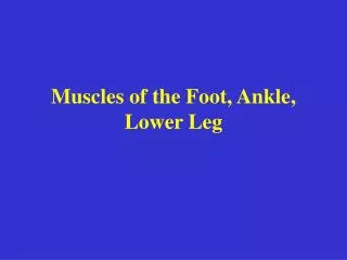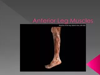
Anterior Leg Muscles
100 likes | 484 Vues
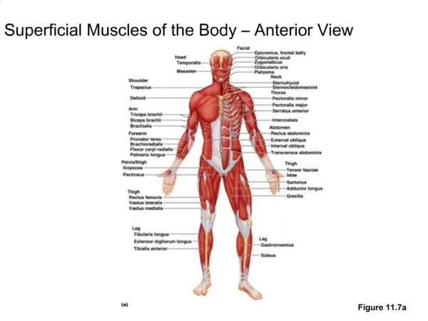
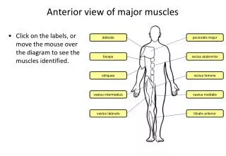
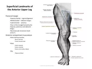
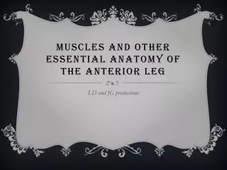
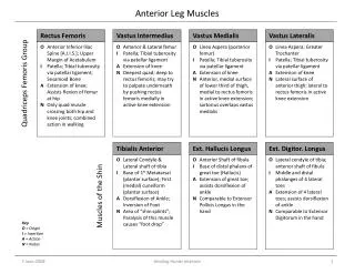
Anterior Leg Muscles. Vastus Lateralis. Rectus Femoris. Ext. Hallucis Longus. Tibialis Anterior. Vastus Intermedius. Ext. Digitor . Longus. Vastus Medialis. Quadriceps Femoris Group. O Linea Aspera ; Greater Trochanter I Patella; Tibial tuberosity via patellar ligament

Anterior Leg Muscles
E N D
Presentation Transcript
Anterior Leg Muscles VastusLateralis Rectus Femoris Ext. HallucisLongus Tibialis Anterior VastusIntermedius Ext. Digitor. Longus VastusMedialis Quadriceps Femoris Group OLinea Aspera; Greater Trochanter IPatella; Tibialtuberosity via patellar ligament AExtension of knee NLateral surface of anterior thigh; lateral to rectus femoris in active knee extension OAnterior & Lateral femur IPatella; Tibialtuberosity via patellar ligament AExtension of knee NDeepest quad; deep to rectus femoris; may try to palpate underneath by pushing rectus femoris medially in active knee extension OLateral condyle of tibia; anterior shaft of fibula IMiddle and distal phalanges of 4 lateral toes AExtension of 4 lateral toes; assists dorsiflexion of ankle NComparable to Extensor Digitorum in the hand OLateral Condyle & Lateral shaft of tibia IBase of 1st Metatarsal (plantar surface); First (medial) cuneiform (plantar surface) ADorsiflexion of Ankle; Inversion of Foot NArea of “shin splints”; Paralysis of this muscle causes “foot drop” OAnterior Shaft of fibula IBase of distal phalanx of great toe (Hallucis) AExtension of great toe; assists dorsiflexion of ankle NComparable to Extensor PollicisLongus in the hand O Anterior Inferior Iliac Spine [A.I.I.S.]; Upper Margin of Acetabulum I Patella; Tibialtuberosity via patellar ligament; Sesamoid Bone A Extension of knee; Assists flexion of femur at hip N Only quad muscle crossing both hip and knee joints; combined action in walking OLinea Aspera (posterior femur) IPatella; Tibialtuberosity via patellar ligament AExtension of knee NAnterior, medial surface of lower third of thigh, medial to rectus femoris in active knee extension; sartorius overlaps vastusmedialis Muscles of the Shin Key O = Origin I = Insertion A = Action N = Notes Healing Hands Institute
Posterior Leg Muscles Hamstrings Notes All three of the Hamstrings cross both the hip and knee joints from lateral to medial (BTM): Biceps Femoris SemiTendinosus SemiMembranosus An inability to touch your toes while keeping your knees extended is largely due to shortened hamstrings. Biceps Femoris Semitendinosus Tibialis Posterior Gastrocnemius Soleus Semimembranosus OPosterior tibia & posterior fibula INavicular; adjacent tarsals & metatarsals on plantar surface of foot AInversion of foot; assists plantarflexion of ankle NBelly of muscle deep to Triceps Surae, cannot be palpated OSoleal line of tibia; posterior head & upper shaft of fibula ICalcaneusvia Achilles Tendon APlantarflexion of ankle (stronger than gastroc.) N“Soleus” (Latin = sole, a flat fish); deep to gastrocnemius; together, gastroc and soleus are often referred to as the “Triceps Surae” OIschialTuberosity IAnterior proximal tibial shaft (inserts medially at the knee, at “PesAnserinus”) AFlexion of Knee;(to a lesser degree, Extension of Hip); Medial rotation of flexed knee NCentral hamstring; tendon is deep, and difficult to palpate OMedial Head: medial epicondyle of femurLateral Head: lateral epicondyle of femur ICalcaneus via Achilles Tendon APlantarflexion of ankle; assists flexion of knee N“Gastro” (Greek = belly); Can act on the knee or the ankle separately, but not simultaneously; raises heel during running & jumping OIschialTuberosity IHead of Fibula (inserts laterally at the knee) AFlexion of Knee;(to a lesser degree, Extension of Hip) NMost lateral of the hamstrings; “biceps” indicates “2 heads” OIschialTuberosity IPosterior medial tibialcondyle AFlexion of Knee;(to a lesser degree, Extension of Hip); Medial rotation of flexed knee NMost medial of the hamstrings; adjacent to Gracilis Hamstrings Muscles of the Calf Key O = Origin I = Insertion A = Action N = Notes Healing Hands Institute
Muscles of the Gluteal Region Gluteus Maximus Gluteus Medius Piriformis Tensor Fasciae Latae Gluteus Minimus OIliac Crest IGreater Trochanter of Femur AAbduction NWhen standing on one foot, Medius contracts on that side to stabilize pelvis and prevent tilting to unsupported side; alternate contraction of these muscles occurs in walking OAnterior sacrum IGreater Trochanter ALateral rotation of femur at hip N(Sciatic Nerve); attempt to palpate just posterior to greater trochanter during active lateral rotation of hip; difficult to differentiate from gluteus medius OIliac Crest (posterior to A.S.I.S.) IIliotibial Tract (I.T. Band) AStabilizes knee; prevents collapse of extended knee during walking NBraces the knee while walking OPosterior sacrum; Ilium; superior gluteal line of ilium IGlutealTuberosity of femur; I.T. Tract AExtension of femur at hip; lateral rotation of extended hip N“Gluteus” (Greek = Rump); Maximus used mostly for power, as in climbing stairs, running, rising from sitting position OPosterior Ilium IAnterior surface of Greater Trochanter AAbduction NGluteus Minimus works with anterior portion of Gluteus Medius Gluteals Deep Lateral Hip Rotator Key O = Origin I = Insertion A = Action N = Notes Healing Hands Institute
Muscles of the Medial Thigh Pectineus Sartorius Gracilis Adductor Magnus Adductor Longus Adductor Brevis OAnterior Pubis ILinea Aspera AAdduction of femur at hip; assists flexion of femur at hip; medial rotation of femur at hip NForms medial border of femoral triangle OInferior Pubic Ramus, Ischialtuberosity & ramus of ischium ILinea Aspera A Adduction of femur at hip; assists flexion & extension of femur at hip NLargest and deepest adductor OAnterior Superior Iliac Spine (A.S.I.S.) IUpper medial shaft of tibia (“PesAnserinus”) AAssists flexion, abduction, lateral rotation of femur at hip; assists flexion, medial rotation of knee NLongest muscle in the body; most superficial thigh muscle; not an adductor OAnterior Pubis IMedial proximal tibia (“PesAnserinus”) AAdduction of femur at hip; assists flexion & medial rotation of flexed knee NMost superficial and medial of adductor group; only adductor that crosses the knee joint; Femur and Gracilis form the shape of the letter “V” OAnterior Pubis ILinea Aspera AFlexion of femur at hip; assists adduction of femur at hip NUppermost of the medial thigh muscles; only adductor that flexes hip O Anterior Pubis I Linea Aspera AAdduction of femur at hip; assists flexion of femur at hip; medial rotation of femur at hip N Not present in all individuals; if present, lies deep to adductor longus Adductors PESANSERINUS Three thigh muscles insert at the Superior (proximal) Medial Tibia forming the shape of a “duck foot”: Sartorius Gracilis Semitendinosus The initial letters of these muscles form the mnemonic expression “Silly Goose Steps”. Muscle of Anterior Thigh Key O = Origin I = Insertion A = Action N = Notes Healing Hands Institute
Muscles of the Lateral Lower Leg Peroneals Notes Peroneus = Greek for “fibula” These muscles would be involved in the case of a lateral ankle sprain. “Eversion” occurs when the foot is turned or rotated outward; i.e., while standing, lifting the lateral edges of the feet while ‘collapsing’ toward the inner arches. PeroneusLongus PeroneusBrevis PeroneusTertius OLateral shaft of fibula IBase of 5th metatarsal AEversion of foot; assists plantar flexion of ankle NHelps when walking or running on uneven surfaces;a.k.a. FibularisBrevis OHead & lateral shaft of fibula IBase of first metatarsal; 1st (medial) cuneiform (plantar surface of foot) AEversion of foot; assists plantar flexion of ankle NTraverses the sole of the foot to meet the tibialis anterior tendon to form a stirrup for the foot (wraps under the foot); a.k.a. FibularisLongus OAnterior distal fibula IBase of 5th metatarsal AEversion of foot; assists dorsiflexion (lifts little toe) NFunctions to place the foot flat on the ground by raising its lateral border Peroneals Key O = Origin I = Insertion A = Action N = Notes Healing Hands Institute