Download
1 / 12
120 likes | 391 Vues
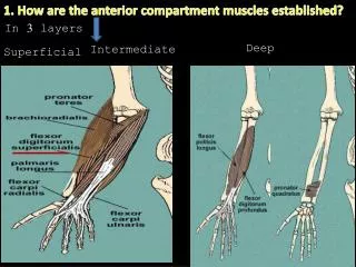
1. How are the anterior compartment muscles established?. In 3 layers . Deep . Intermediate. Superficial. 2. ...the muscles of the superficial layer?. Superficial layer flexor carpi radialis flexor carpi ulnaris palmaris longus pronator teres. Medial epicondyle of humerus.

E N D
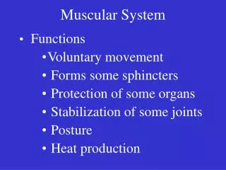
1. How are the anterior compartment muscles established? In3 layers Deep Intermediate Superficial
2. ...the muscles of the superficial layer? Superficial layer flexor carpi radialis flexor carpi ulnaris palmaris longus pronator teres Medial epicondyle of humerus Pisiform
3. ..the muscles of the intermediate and deep layers? Intermediate layer flexor digitorum superficialis Deep layer flexor digitorum profundus pronator quadratus flexor pollicis longus Middle phalanx - shaft Distal phalanx-base Distal phalanx -base
4. How is the arterial supply of the anterior compartment? Inf. border of teres major Axillary artery Till where? Brachial artery Bracihal artery divides into Ulnar artery (medial) Radial artery (lateral) Neck of the radius
5. Which veins do you see in the anterior compartment? (Paired) Deep veins accompanying veins plentiful in the forearm. arise from the anastomosing deep venous palmar arch in the hand. drain into brachial veins in the cubital fossa.
6. ...the median nerve in the anterior compartment? principal nerve no branches in the arm other than small twigs to the brachial artery. Its major branch in the forearm anterior interosseousnerve Leaves the cubital fossa by passing between 2 heads of the pronator teres and humero-ulnar and radial heads of the flexor digitorum superficialis.
7. ...the ulnar nerve in the anterior compartment? Enters the anterior compartment by passing posteriorly around medial epicondyle of humerus and between humeral and ulnar heads of flexor carpi ulnaris muscle Two small cutaneous branches; palmar branch & dorsal branch
8. ....the radial nerve in the anterior compartment? motor and sensory functions in both arm & forearm (but only sensory functions in the hand) in the forearm Superficial (sensory) deep to brachioradialis deep (motor) between two heads of supinator
9. What are the boundaries of the cubital fossa? • Superiorly imaginary line connecting medial &lateral epicondyles. • Mediallypronator teres. • Laterallybrachioradialis.
10. ...the contents of the cubital fossa? • 1) Terminal part of the brachial artery,radial and ulnar arteries • 2) Biceps brachii tendon • 3) Median nerve • 4) Radial nerve • 5) (Deep) accompanying veins of the arteries
11...the contents of the cubital fossa? Superficially, in the subcutaneous tissue overlying the fossa median cubital vein, medial and lateral antebrachial cutaneous nerves basilic and cephalic veins.
12. ....nerves in the cubital fossa? median nervelies immediately medial to the brachial artery and leaves the fossa by passing between the ulnar and humeral heads of the pronator teres muscle. radial nervelies under brachioradialis (lateral margin of the fossa) gives off deep branch of the radial nerve and continues as superficial radial nerve.