Download

1 / 60
600 likes | 788 Vues
University of Bologna Prof. Marco Leonardi Neuroradiology Department Bellaria Hospital Bologna. Considerations on Advanced MRI Techniques in studying brain gliomas. A.Bacci, F. Menetti, P. Agati, R. Agati, M. Messia, S. Ascanio, M. Leonardi.

E N D
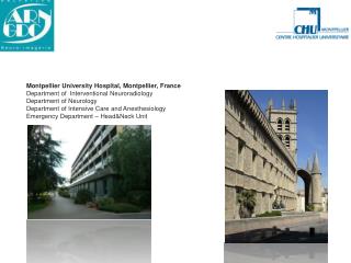
University of Bologna Prof. Marco Leonardi Neuroradiology Department Bellaria Hospital Bologna
Considerations on Advanced MRI Techniques in studying brain gliomas. A.Bacci, F. Menetti, P. Agati, R. Agati, M. Messia, S. Ascanio, M. Leonardi
Advanced MR Techniques • Since 2004 we have used our 3T equipment and the advanced MR Techniques (DWI, PWI, SV Spectroscopy) in studying brain tumours • Our 3T experience: a careful selection, setting up and optimization of the sequences used in order to avoid unsatisfactory results is mandatory • Could advanced MR Techniques increase the proportion of correctly diagnosed cases?
3T 1.5T • Higher S/N ratio • Longer T1, shorter T2* and T2; • Reduction of r1 relaxivity (contrast media) • Increase of magnetic susceptibility • Increase of geometric distortion • “Chemical Shift” increase • More flow artifacts • More SAR • More acoustic noise • Increase of dieletric effect
3T 1.5T • Higher spatial resolution • Higher temporal resolution • “Complex” studies • Different approach to the sequences • Pathologic Enhancement (SE, Flair or SPGR?): T1 • Morphology: T2 (no gap) • Careful selection, setting up and optimization of the sequences used in order to avoidunsatisfactory results
3T 1.5T • Higher spatial resolution • Higher temporal resolution • “Complex” studies Different approach to the sequences Pathologic Enhancement (SE, Flair or SPGR?): T1 Morphology: T2 (no gap) Careful selection, setting up and optimization of the sequences used in order to avoid unsatisfactory results
3T 1.5T Higher spatial resolution Higher temporal resolution “Complex” studies • Different approach to the sequences • Pathologic Enhancement (SE, Flair or SPGR?): T1 • Morphology: T2 (no gap) • Careful selection, setting up and optimization of the sequences used, in order to avoidunsatisfying results
When 3T? • Intraaxial Tumours • Microadenomas • Epilepsy • White matter diseases • Angio-MR (Tricks, Spine..) • fMRI
Tumour Protocol 8 channels phased array coil • 1H-MRS SV (TR 2000, TE 35) • PWI (SE-EPI) • Sag SE T1-w CE • Ax SE T1-w CE • Cor SE T1-w CE • Sag SE T1-w • Ax Flair T2-w • Ax FRFSE T2-w • Ax SE T1-w • DWI
Tumour Protocol 8 channels phased array coil • 1H-MRS SV (TR 2000, TE 35) • PWI (SE-EPI) • Sag SE T1-w CE • Ax SE T1-w CE • Cor SE T1-w CE • Sag SE T1-w • Ax Flair T2-w • Ax FRFSE T2-w • Ax SE T1-w • DWI
SE T1-w on 3T?(Pathologic enhancement) • On 3T, Flair or SPGR T1-w sequences allow a better grey-white matter contrast than SE • However we have decided to use SE (as 1.5T) because of the improved sensitivity at: • pathologic enhancement after contrast media administration • haemoglobin degradation products
SPGR e/o Flair T1-w vs SE Flair SE SPGR
SE SPGR
PWI • It is well known that a GE-EPI sequence is more sensitive to the first pass of gadolinium than a SE sequence • However we decided to use a SE-EPI sequence because of: • Less sensitivity to magnetic susceptibility artifacts along the cranial theca • Improved sensitivity only to contrast agent within the capillaries (i.e. neoangiogenesis) (S.Cha et coll. Radiology 2002; 223:11-29)
Magnetic susceptibility artifacts GE-EPI SE-EPI
The first pass of gadolinium(Signal decreases) SE-EPI GE-EPI
Spectroscopy Single voxel, TR 2000, TE 35 • better S/N ratio and spectral resolution • quantification of variation of the metabolites more than spatial distribution of metabolites (as in CSI) • Relatively long acquisition time (5 min.)
Equipment(Complex studies) • 2 Workstations RIS-PACS • 2 Workstations ADW • 2 Functool • 1 Brainwave • 1 Sage • 1 PC Windows (BrainVoyager) • 1 PC Windows (Access) • 1 PC Windows • Presentation/Stim 2 • Hardware for activation studies • 1 PC Linux (LC-Model)
Work (still) in progress… For example we are now evaluating: • other software to elaborate PWI studies such as Brainstat and DPTools (correction of the T1 effect) • Arterial Spin labelling • High Order Shimming (Spectroscopy)
Use of advanced MR Techniques(Our experience) • Individual cases • More systematic way, in population studies • Glioma grading • Recurrent/residual tumours vs treatment related changes (e.g. radionecrosis)
Use of advanced MR Techniques(Our experience) • Individual cases More sistematic way, in population studies Glioma grading Recurrent/residual tumor vs treatment related changes
Diffusion (High cellularity)
Perfusion (High Neoangiogenesis)
Spectroscopy (Intralesional Necrosis)
Lip Cho NAA ????
Lip Cho Cr NAA mI Radiation injury or recurrent tumour?
Lip Cho NAA Cr mI Recurrenttumour 17/07/08 17/07/08
Use of advanced MR Techniques(Our experience) Single cases • More systematic way, in population studies • Glioma grading • Recurrent/residual tumour vs treatment related changes
Glioma grading • MRI is capable of providing important morphological information but it is not very accurate in tumour grading (20-30% of gliomas are misclassified) * • Could DWI, PWI and 1H-MRS studies increase the proportion of correctly diagnosed cases? • Is it possible to define threshold values? *Neuroradiology 2002; 44:371-381 Radiology 1990;174:411-415 Radiology 1999:211:791-798 Neuroradiology 1992; 34:463-469
MRI in gliomas Li X et al. JMRI 16:229-237, 2002
MRI in gliomas • Addition of 1H-MRS and perfusion information to MRI studies significantly increases the proportion of correctly diagnosed cases • Sensitivity, specificity, PPV and NPV to differentiate between high and low-grade gliomas (160 patients) with: • MRI: 72.5%, 65%, 86.1%, 44.1% • rCBV+Cho/Cr+Cho/NAA: 93.3%, 60%, 87.5%, 75.0% Law M et al. AJNR 24:1989-98, 2003
Our Study • Grade II (n=25) • 11 Astrocytomas • 12 Oligodendrogliomas • 2 Oligoastrocytomas • Grade III (n=21) • 11 Anaplastic Astrocytomas • 5 Anaplastic Oligodendrogliomas • 4 Anaplastic Oligoastrocytomas • 1 Gliomatosis • Grade IV (n=31) : GBM
Our experience All patients underwent preoperative MR examination on a 3T system: MRI DWI (ADC ratio) PWI (rCBV ratio) SV MRS Histopathologic evaluation
Statistical analysis (Test Brunner Dette Monk)
Evidence of statistically significant differences between two classes (i.e. grade II and grade III+IV gliomas) is of limited practical utility due to overlapping values between different glioma grades • Our aim should be to identify threshold values to define the grade of a glioma in individual cases
Cart Software • It is a decision tree that automatically sifts large, complex databases, searching for and isolating significant patterns and relationships. • CART can reveal important data relationships (and threshold values) that could remain hidden using other analytical tools.
ADC min 1.09 rCBV max 2.02 mI/Cre 1.36 Grade II vs III+IVMRI and Advanced Techniques Lac/Cre 3.45