Download
1 / 1
10 likes | 14 Vues
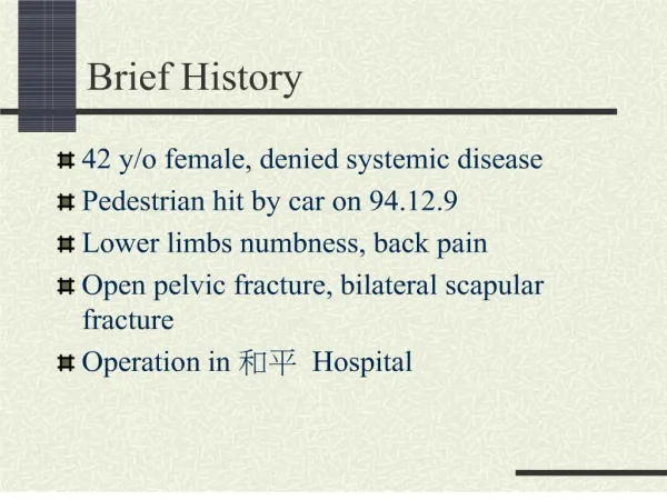
Extra-peritoneal Packing of Hemorrhagic Pelvic Fracture By: Thomas S. Kefalas & Colby DeCapua Lock Haven University Physician Assistant Program. Figure 4. Extra- peritoneal pelvic packing (1). Figure 1. Fractured pelvis (5). Studies

E N D
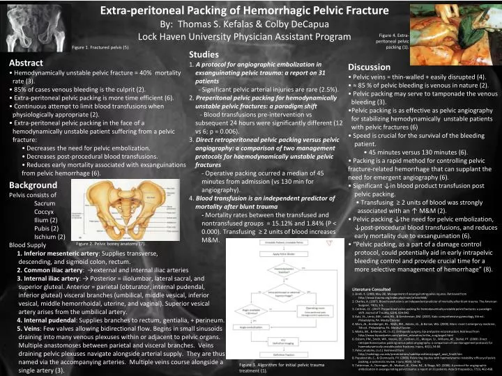
Extra-peritoneal Packing of Hemorrhagic Pelvic Fracture By: Thomas S. Kefalas & Colby DeCapua Lock Haven University Physician Assistant Program Figure 4. Extra- peritoneal pelvic packing (1). Figure 1. Fractured pelvis (5). • Studies • 1. A protocol for angiographic embolization in exsanguinating pelvic trauma: a report on 31 patients • - Significant pelvic arterial injuries are rare (2.5%). • 2. Preperitonal pelvic packing for hemodynamically unstable pelvic fractures: a paradigm shift • - Blood transfusions pre-intervention vs subsequent 24 hours were significantly different (12 vs 6; p = 0.006). • 3. Direct retroperitoneal pelvic packing versus pelvic angiography: a comparison of two management protocols for haemodynamically unstable pelvic fractures • - Operative packing ocurred a median of 45 minutes from admission (vs 130 min for angiography). • 4. Blood transfusion is an independent predictor of mortality after blunt trauma • - Mortality rates between the transfused and nontransfused groups = 15.12% and 1.84% (P < 0.000). Transfusing ≥ 2 units of blood increases M&M. • Abstract • • Hemodynamically unstable pelvic fracture = 40% mortality rate (3). • • 85% of cases venous bleeding is the culprit (2). • • Extra-peritoneal pelvic packing is more time efficient (6). • • Continuous attempt to limit blood transfusions when physiologically appropriate (2). • • Extra-peritoneal pelvic packing in the face of a hemodynamically unstable patient suffering from a pelvic fracture: • • Decreases the need for pelvic embolization. • • Decreases post-procedural blood transfusions. • • Reduces early mortality associated with exsanguinations from pelvic hemorrhage (6). • Discussion • • Pelvic veins = thin-walled + easily disrupted (4). • • ≈ 85 % of pelvic bleeding is venous in nature (2). • • Pelvic packing may serve to tamponade the venous bleeding (3). • •Pelvic packing is as effective as pelvic angiography for stabilizing hemodynamically unstable patients with pelvic fractures (6) • • Speed is crucial for the survival of the bleeding patient. • • 45 minutes versus 130 minutes (6). • • Packing is a rapid method for controlling pelvic fracture-related hemorrhage that can supplant the need for emergent angiography (6). • • Significant ↓in blood product transfusion post pelvic packing. • • Transfusing ≥ 2 units of blood was strongly associated with an ↑ M&M (2). • • Pelvic packing ↓the need for pelvic embolization, ↓post-procedural blood transfusions, and reduces early mortality due to exsanguination (6). • • “Pelvic packing, as a part of a damage control protocol, could potentially aid in early intrapelvic bleeding control and provide crucial time for a more selective management of hemorrhage” (8). • Background • Pelvis consists of • Sacrum • Coccyx • Ilium (2) • Pubis (2) • Ischium (2) • Blood Supply • 1. Inferior mesenteric artery: Supplies transverse, • descending, and sigmoid colon, rectum. • 2. Common iliac artery: →external and internal iliac arteries • 3. Internal iliac artery: → Posterior = iliolumbar, lateral sacral, and superior gluteal. Anterior = parietal (obturator, internal pudendal, • inferior gluteal) visceral branches (umbilical, middle vesical, inferior vesical, middle hemorrhoidal, uterine, and vaginal). Superior vesical • artery arises from the umbilical artery. • 4. Internal pudendal: Supplies branches to rectum, gentialia, + perineum. • 5. Veins: Few valves allowing bidirectional flow. Begins in small sinusoids draining into many venous plexuses within or adjacent to pelvic organs. Multiple anastomoses between parietal and visceral branches. Veins draining pelvic plexuses navigate alongside arterial supply. They are thus named via the accompanying arteries. Multiple veins course alongside a single artery (3). Figure 2. Pelvic boney anatomy (7). Literature Consulted 1. Brohi, K. (2008, May 20). Management of exsanguinating pelvis injuries. Retrieved from http://www.trauma.org/index.php/main/article/668/ 2. Charles, A. (2007). Blood transfusion is an independent predictor of mortality after blunt trauma. The American Surgeon, 73(1), 1-5. 3. Cothren, CC. (2007). Preperitonal pelvic packing for hemodynamically unstable pelvic fractures: a paradigm shift. Journal of Trauma, 62(4), 834-842. 3. Katz, VL., Lentz, GM., Lobo, RA., & Gershenson, DM. (2007). Katz: comprehensive gynecology, 5th ed.. Philadelphia, PA: Mosby Elsevier. 4. Marx, JA., Hockberger, RS., Walls, RM., Adams, JG., & Barsan, WG. (2009). Marx: rosen's emergency medicine, 7th ed.. Philadelphia, PA: Mosby Elsevier. 5. Matta, JM., & Klenck, RE. (n.d.). Orthopedic surgery, hip and pelvis reconstruction. Retrieved from http://www.hipandpelvis.com/patient_education/ortho_surg/page2.html 6. Osborn, PM., Smith, WR., Moore, EE., Cothren, CC., Morgan, SJ., Williams, AE., Stahel, PF. (2009). Direct retroperitoneal pelvic packing versus pelvic angiography: a comparison of two management protocols for haemodynamically unstable pelvic fractures. Injury, 40(1), 54-60. 7. Pelvic anatomy. (n.d.). Retrieved from http://radiology.usc.edu/presentations/saddleprosthesis/page2_anat_final2.htm 8. Popakostidis, C., & Giannoudis, PV. (2009). Pelvic ring injuries with haemodynamic instability: efficacy of pelvic packing, a systematic review. Injury, 40(4), 53-61. 9. Totterman, A., Dormagen, JB., Madsen, JE., Klow, NE., & Skaga, NO. (2006). A protocol for angiographic embolization in exsanguinating pelvic trauma: a report on 31 patients. ActaOrthopaedica, 77(3), 462-468. Figure 3. Algorithm for initial pelvic trauma treatment (1).