Download
1 / 36
360 likes | 385 Vues
Explore the intricate structures within the cell's cytoplasm, including organelles like ribosomes, endoplasmic reticulum, Golgi apparatus, lysosomes, and mitochondria. Learn about the role of the cytoskeleton, centrosomes, and cell diversity.

E N D
PART 2 Cells: The Living Units
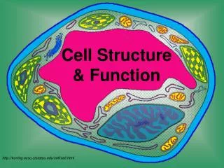
The Cytoplasm • Cytoplasm – lies internal to plasma membrane • Consists of cytosol, organelles, and inclusions • Cytosol (cytoplasmic matrix) • Jelly-like fluid in which other cellular elements are suspended • Consists of water, ions, and enzymes
Cytoplasmic Organelles • Ribosomes – constructed of proteins and ribosomal RNA • Site of protein synthesis • NOTE: Most antibiotics work by blocking bacterial protein synthesis. The antibiotic works on one of the subunits to prevent bacteria from multiplying!
Structure of a Generalized Cell Figure 2.1
Cytoplasmic Organelles • Endoplasmic reticulum – “network within the cytoplasm” • Rough ER – ribosomes stud the external surfaces • Smooth ER – consists of tubules in a branching network • No ribosomes are attached; therefore no protein synthesis
The Endoplasmic Reticulum and Ribosomes Figure 2.5
Assembly of Proteins at the Rough Endoplasmic Reticulum Figure 2.6
Cytoplasmic Organelles • Golgi apparatus – a stack of three to 10 disk-shaped envelopes • Sorts products of rough ER and sends them to proper destination • Products of rough ER move through the Golgi from the convex (cis) to the concave (trans) side
Role of the Golgi Apparatus in Packaging Products of Rough ER Figure 2.8
Cytoplasmic Organelles • Lysosomes – membrane-walled sacs containing digestive enzymes • Digest unwanted substances • Peroxisomes – membrane-walled sacs of oxidase enzymes • Enzymes neutralize free radicals and break down poisons • Break down long chains of fatty acids • Are numerous in the liver and kidneys
Mitochondria • Mitochondria – generate most of the cell’s energy; most complex organelle • More abundant in energy-requiring cells, like muscle cells and sperm Figure 2.10
Cytoplasmic Organelles • Cytoskeleton – “cell skeleton” – an elaborate network of rods • Contains three types of rods • Microtubules – cylindrical structures made of proteins • Microfilaments – filaments of contractile protein actin • Intermediate filaments – protein fibers
Cytoskeleton: Microtubule Figure 2.11a
Cytoskeleton: Microfilament Figure 2.11b
Cytoskeleton: Intermediate Filament Figure 2.11c
Cytoplasmic Organelles • Centrosomes and centrioles • Centrosome – a spherical structure in the cytoplasm • Composed of centrosome matrix and centrioles • Centrioles – paired cylindrical bodies • Consists of 27 short microtubules • Act in forming cilia • Necessary for karyokinesis (nuclear division)
Cytoplasmic Inclusions • Temporary structures • Not present in all cell types • May consist of pigments, crystals of protein, and food stores • Lipid droplets – found in liver cell and fat cells • Glycosomes – store sugar in the form of glycogen
The Nucleus • The nucleus – “central core” or “kernel” – control center of cell • DNA directs the cell’s activities • Nucleus is approximate 5µm in diameter
The Nucleus Figure 2.13
The Nucleus • Nuclear envelope – two parallel membranes separated by fluid-filled space • Chromatin – composed of DNA and histone proteins • Condensed chromatin – contains tightly coiled strands of DNA
The Nucleus • Chromatin – composed of DNA and histone proteins • Extended chromatin – contains uncoiled strands of DNA • DNA’s genetic code is copied onto mRNA (transcription) while in “extended chromatin” form • Chromosomes – highest level of organization of chromatin • Contains a long molecule of DNA
The Nucleus • Nucleolus – “little nucleus” – in the center of the nucleus • Contains parts of several chromosomes • Site of ribosome subunit manufacture
Cellular Diversity • Specialized functions of cells relates to • Shape of cell • Arrangement of organelles
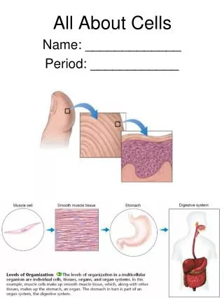
Cellular Diversity • Cells that connect body parts or cover organs • Fibroblast – makes and secretes protein component of fibers • Erythrocyte – concave shape provides surface area for uptake of the respiratory gases • Epithelial cell – hexagonal shape allows maximum number of epithelial cells to pack together
Cells that Connect Body Parts or Cover Organs Figure 2.16, step 1
Cellular Diversity • Cells that move organs and body parts • Skeletal and smooth muscle cells • Elongated and filled with actin and myosin • Contract forcefully
Cells that Move Organs and Body Parts Figure 2.16, step 2
Cellular Diversity • Cells that store nutrients • Fat cell – shape is produced by large fat droplet in its cytoplasm • Cells that fight disease • Macrophage – moves through tissue to reach infection sites
Cells that Store Nutrients and Cells that Fight Disease Figure 2.16, steps 3–4
Cellular Diversity • Cells that gather information • Neuron – has long processes for receiving and transmitting messages Figure 2.16, step 5
Cellular Diversity • Cells of reproduction • Oocyte (female) – largest cell in the body • Contains many copies of organelles for distribution to daughter cells • Sperm (male) – possesses long tail for swimming to the egg for fertilization Figure 2.16, step 6
The Cell Life Cycle • Is the series of changes a cell goes through • Interphase • G1 phase – growth 1 or Gap 1 phase • The first part of interphase • Cell metabolically active – growth – make proteins • Variable in length from hours to YEARS (egg cell) • Centrioles begin to replicate near the end of G1 PLAY Late Interphase
The Cell Life Cycle • S (synthetic) phase – DNA replicates itself • Ensures that daughter cells receive identical copies of the genetic material (chromatin extended) • G2 phase – growth 2 or Gap 2 • Centrioles finish copying themselves • Enzymes needed for cell division are synthesized in G2 • During S (synthetic) and G2 phases – cell carries on normal activities
The Cell Life Cycle Figure 2.17
The Cell Life Cycle • Cell division • M (mitotic) phase – cells divide during this stage • Follows interphase (G1, S, and G2)
The Cell Life Cycle • Cell division involves • Mitosis – division of the nucleus during cell division • Chromosomes are distributed to the two daughter nuclei • Cytokinesis – division of the cytoplasm • Occurs after the nucleus divides PLAY Mitosis Overview