Download

1 / 37
370 likes | 511 Vues
neoplasia III tumour genetics MOLECULAR B ASIS OF CANCER. 1. Nonlethal genetic damage lies at the heart of carcinogenesis 2. Tumor is formed by the clonal expansion of a single precursor cell

E N D
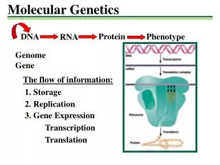
1. Nonlethal genetic damage lies at the heart of carcinogenesis 2. Tumor is formed by the clonal expansion of a single precursor cell 3. The principal targets of genetic damage are the 4 classes of normal regulatory genes 4. Carcinogenesis is a multistep process resulting from accumulation of multiple mutations FUNDAMENTAL PRINCIPLES of carcinogenesis
four classes of normal regulatory genes • Growth-promoting - proto-oncogenes • Growth-inhibiting - tumor suppressor genes • Genes that - regulate apoptosis • Genes involved in - DNA repair
Proto- oncogenes- mutant alleles are dominant Tumor suppressor genes - both normal alleles must be damaged to transform cell Genes regulating apoptosis - may behave like proto-oncogenes or tumor supressor genes DNA repair genes - affect indirectly influence the ability to repair nonlethal damage in other genes fundamental principles
Tumor progression and heterogeneity result from multiple mutations that accumulate independently in diff cells generating subclones with varying abilities to grow, invade, metastasize and resist /respond to therapy
7 Key changes in cell physiology for malignant transformation : Self-sufficiency in growth signals Insensitivity to growth-inhibitory signals Evasion of apoptosis Limitless replication potential Sustained angiogenesis Ability to invade and metastasize Defects in DNA repair
Proto-oncogenes – normal cellular genes , multiple roles participating in cellular functions related to growth and proliferation • Poteins encoded by proto-oncogenes may act as : • Growth factors • Growth factor receptors • Signal transducers • Transcription factors • Cell cycle components CaNCER – ASSOCIATED GENES
Oncognes – genes that promote autonomous cell growth in cancer cells • Created by mutations in proto-oncogenes - constitutively active oncogenes • Oncoproteins – produts of oncogenes , resemble products of proto-oncogene except • devoid of imp internal regulatory elements • Endow the cell with self-sufficiency in growth
The binding of a growth factor to its specific receptor • Transient & limited activation of the growth factor receptor, in turn activates signal-transducing proteins on the inner leaflet of the plasma membrane The sequential steps of normal cell proliferation :
Transmission of the transduced signal across the cytosol to the nucleus via second messengers or by a cascade of signal transduction molecules Induction and activation of nuclear regulatory factors that initiate DNA transcription Entry and progression of the cell into the cell cycle, ultimately resulting in cell division The sequential steps of normal cell proliferation.
Overview of the main types of cell surface receptors and their principal signal transduction pathways
Signaling from tyrosine kinase receptors. Binding of the growth factor (ligand) causes receptor dimerization and autophosphorylation of tyrosine residues.
Cell cycle landmarks. The figure shows the cell cycle phases (G0, G1, G2, S, and M), the location of the G1 restriction point, and the G1/S and G2/M cell cycle checkpoints
Oncoproteins encoded by oncogenes endow the cell with self-sufficiency in growth. • Now, we will discuss that • What are the functions of oncogene products, the oncoproteins? • How do the normally “civilized” proto-oncogenes turn into “enemies within”? Self-sufficiency in growth signals : Oncogenes
Normal cells require stimulation by growth factors to undergo proliferation. • Most soluble growth factors have paracrinesignaling . growth factors
Cancer cells • Acquire the ability to synthesize the same growth factors to which they are responsive, generating an autocrine loop; e.g • Many glioblastomas secrete platelet-derived growth factor (PDGF) and express the PDGF receptor • Many sarcomas make both transforming growth factor α (TGF-α) and its receptors growth factors
Overexpression of growth factor genes ; forcing cells to secrete large amounts of growth factors • Increased cell proliferation contributes to malignant change by increasing the risk of mutations (spontaneous/induced) growth factors
Selected Oncogenes, Their Mode of Activation, and Associated Human Tumors
Oncogenic versions of receptors have constitutive dimerization and activation without binding to growth factor Mutant receptors deliver continuous mitogenic signals to cells, even in the absence of growth factor These receptors can be constitutively activated in tumors by multiple mechanisms including mutations , gene rearrangements, overexpression c-KIT , HER-2/NEU - Targeted therapy growth factor receptors
Selected Oncogenes, Their Mode of Activation, and Associated Human Tumors
Signal-Transducing Proteins • Several oncoproteins mimic the function of normal cytoplasmic signal-transducing proteins ; strategically located on the inner leaflet of the plasma membrane • Biochemically, the signal-transducing proteins are heterogeneous. • The most well-studied example of a signal-transducingoncoprotein is the RAS family of guanine triphosphate (GTP)-binding proteins (G proteins).
Three RAS genes in humans (HRAS, KRAS, NRAS). • Point mutation of RAS family genes is the single most common abnormality of proto-oncogenes in human tumors • Approximately 15% to 20% of all human tumors contain mutated versions of RAS proteins • Carcinomas colon (50%) and pancreas (90%) have mutations of KRAS, bladder tumors have HRASmutations, and hematopoietic tumors bear NRASmutations. • RAS mutations are infrequent in certain other Cas such as Ca cervix or breast. The RAS Oncogene
. GTP hydrolysis by GTPase converts RAS into inactive form ; this is markedly accelerated by GAPs. Thus GAPs function as “brakes” to prevent uncontrolled activation of RAS. The mutated RAS is trapped in active form because it cannot hydrolyse GTP. The RAS Oncogene Model for action of RAS genes
Mutations that unleash latent oncogenic activity occur in several non-receptor-associated tyrosine kinases, which normally function in signal transduction pathways • Chromosomal translocation or rearrangements that create fusion genes encoding constitutively active tyrosine kinases. • In CML and some acute lymphoblastic leukemias, the ABL gene is translocated from its normal abode on chromosome 9 to chromosome 22, where it fuses with the BCR gene. Alterations in Nonreceptor Tyrosine Kinases
The resultant chimeric gene encodes a constitutively active, oncogenic BCR-ABL tyrosine kinase. • Treatment of CML has been revolutionalized by development of “designer” drug that inhibits BCR-ABL kinase Chromosomal translocation & associated oncogenes
The ultimate consequence of signaling through oncogenes like RAS or ABL is inappropriate and continuous stimulation of nuclear transcription factors that drive growth-promoting genes. • Transcription factors contain specific amino acid sequences or motifs that allow them to bind DNA or to dimerize for DNA binding. • Binding of these proteins to specific sequences in the genomic DNA activates transcription of genes. Transcription Factors
Growth autonomy may occur as a consequence of mutations affecting genes that regulate transcription. • A host of oncoproteins, including products of the MYC, MYB, JUN, FOS, and REL oncogenes, are transcription factors that regulate the expression of growth-promoting genes, such as cyclins. • MYC is the most commonly involved in human tumors. Transcription Factors
MYC protooncogene is expressed in all eukaryotic cells and is rapidly induced when cell gets signal to divide. After transient ↑ of MYC mRNA the expression declines to basal level • MYC interacts with components of the DNA-replication machinery, and plays a role in the selection of origins of replication. • Overexpression of MYC may drive activation of more origins than needed for normal cell division, or bypass checkpoints involved in replication, leading to genomic damage and accumulation of mutations. . The MYC Oncogene
Finally, MYC is one of a handful of transcription factors that can act in concert to reprogram somatic cells into pluripotent stem cells • MYC may also enhance self-renewal, block differentiation, or both. • Amplification of this gene is associated with a variety of tumors, e.gBurkit lymphoma The MYC Oncogene
Amplification of the N-MYC gene in human neuroblastomas. The N-MYC gene, normally present on chromosome 2p, becomes amplified and is seen either as extra chromosomal double minutes or as a chromosomally integrated, homogeneous staining region (HSR). The integration involves other autosomes, such as 4, 9, or 13. The MYC Oncogene
The orderly progression of cells through various phases of cell cycle is orchestrated by cyclin-dependent kinases –CDKs , which are activated by binding to cyclins ;( cyclin D,E ,A & B appear sequentially); • CDK- cyclin complexes phosphorylate crucial target proteins that drive the cell through cell cycle ;on completion of this task cyclin levels decline rapidly • Cyclin D genes are overexpressed in many cancers (breast, esophagus , liver, some lymphomas) • Amplification of CDK4 gene occurs in melanomas sarcomas , glioblastomas CYCLIN AND CYCLIN DEPENDENT KINASES
While cyclins activate CDKs, CDK inhibitors (CDKIs) silence CDKs , exert negative effect over the cell cycle Expression of these inhibitors is down regulated by mitogenic signaling pathways, thus promoting progression of cell cycle CDKIs are mutated or silenced in many human malignancies