Esophagus and Diaphragmatic Hernia
290 likes | 901 Vues
Esophagus and Diaphragmatic Hernia. Basic Science Lecture 3/8/11 Marcie Dorlon, PGY3. Esophageal Anatomy. Blood Supply : Inferior thyroid, aortic branches, bronchial branches, left gastic artery branches, inferior phrenic branches

Esophagus and Diaphragmatic Hernia
E N D
Presentation Transcript
Esophagus and Diaphragmatic Hernia Basic Science Lecture 3/8/11 Marcie Dorlon, PGY3
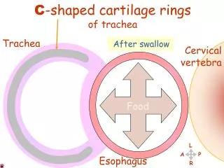
Esophageal Anatomy Blood Supply: Inferior thyroid, aortic branches, bronchial branches, left gastic artery branches, inferior phrenic branches Venous drainage: inferior thyroid vein, bronchial veins, hemiazygous and azygous veins, coronary vein Lymphatic Drainage: submucosal, dense single-plexus Innervation: mainly vagus
Assessment of Esophageal Function • Antireflux mechanisms: • mechanically effective LES • efficient esophageal clearance • adequately functioning gastric reservoir • Tests to detect: • structural abnormalities • functional abnormalities • increased exposure to gastric juice • duodenogastric function as related to esophageal disease
Esophageal Studies • Radiographic evaluation (barium swallow) • endoscopic evaluation (with or without biopsy) • stationary manometry • high-resolution manometry • esophageal impedance • esophageal transit scintigraphy • video- and cineradiography • 24-hour ambulatory pH monitoring • gastric emptying study • gastric acid analysis • cholescintigraphy • 24-hour gastric pH monitoring
GERD • Chronic disease often requiring life-long medical tx • Dx: Lack of universally accepted definition • Sx: heartburn (substernal burning worsened by spicy and fatty foods, etoh, coffee, chocolate, etc) and regurgitation, cough, hoarseness, asthma, recurrent pneumonia, bronchospasm, dysphagia, odynophagia • Complications: esophagitis, stricture, Barrett’s esophagus (metaplasia), adenocarcinoma (neoplasia), repetitive aspiration and pulmonary fibrosis • Tx first line: H2 blockers, PPI, surgery if medical management fails • Surgical Tx Indicated: Barrett’s metaplasia, ulcer, stricture, failure of medical tx, structurally defective LES • Anti-reflux surgery restores gastroesophageal barrier • Laparoscopic Nissen Fundoplication, TransthoracicNissen Fundoplication
Giant Diaphragmatic (Hiatal) Hernias • Type I sliding hernia, upward dislocation of cardia in the posterior mediastinum • Type II rolling/paraesophageal hernia, upward dislocation of gastric fundus alongside normally positioned cardia • Type III combined sliding-rolling/mixed hernia, upward dislocation of cardia and gastric fundus • Type IV: additional organ (colon) herniates along with stomach into chest • Sx: same as GERD, anemia • Dx: Barrium swallow, endoscopy • Indications for surgery: paraesophageal hernia (prevent bleeding, infarction, perforation) • Approach: transabdominal or transthoracic to repair diaphragm +/- fundoplication • Recurrence rates 10-15%
Schatzki’s Ring • Thin circumferential submucosal ring in the lower esophagus at the squamocolumnar junction, often associated with hiatal hernia • Significance and pathogenesis unclear, prevalent in 0.2-14% of population • Symptoms: dysphagia (solid food), reflux • Tx: dilation, incision or excision of ring, antireflux procedure
Scleroderma • Esophageal abnormalities in 80% patients with this diagnosis • Perivascular deposition of collagen leads to smooth muscle atrophy in GI tract • Primary neurogenic disorder in regards to esophageal symptoms as methacholine and edrophonim relieve symptoms • Dx: manometry demonstrates absence of peristalsis in distal smooth muscle portion, progressively weakened LES • Can lead to strictures and severe esophagitis • Tx: medical, serial dilations, partial fundoplication for severe cases
Motility Disorders • : Zenker’sdiverticulum- most common sign of pharyngoesophageal dysfunction • Sx: dysphagia, regurgitation of undigested bland material, chronic aspiration, weight loss • Dx: barium swallow • Tx: open cricopharyngealmyotomy, diverticularpexy, diverticulectomy, endoscopic • Achalasia- primary disorder of LES not relaxing • Sx: pain, regurgitation, weight loss • Dx: 24-Hr motility monitoring, radiographic “bird’s beak” narrowing distal esophagus • Tx: dilation, medication, Botox injection, surgery • Diffuse esophageal spasm (DES), nutcracker esophagus, hypertensive LES
Operations for Motor Disorders of Esophagus • Long Myotomy- thoracic approach, myotomy extends from 1-2cm below GEJ proximally to level of dysmotility • Heller Myotomy- myotomy of LES via thoracic or abdominal approach • Open Esophageal Myotomy- used for reoperation • Laparoscopic Heller Myotomy and partial fundoplication- beat pneumatic dilation and Botox injections in several RCT for esophageal motility disorders
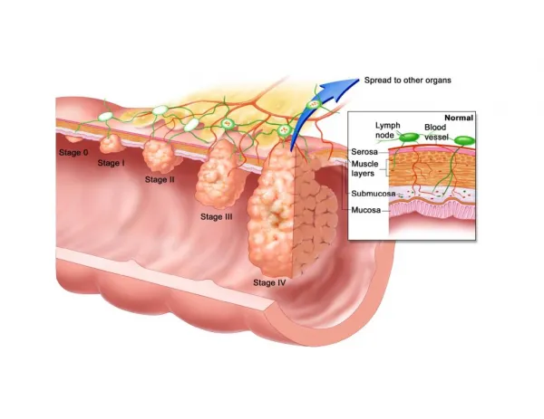
Carcinoma of Esophagus • Most common worldwide: squamous carcinoma, associated with etoh and tobacco, lye ingestion, long standing achalasia, HPV • Increasing incidence of adenocarcinoma: associated with GERD and Barrett’s esophagus • Sx: Dysphagia, asymptomatic found in EGD, stridor, cough, choking, recurrent aspiration or pneumonia, pain swallowing rough or dry food , vocal cord paralysis, TEF • Staging by CXR, CT, PET, EUS • Tx: chemoradiation and surgery
Sarcoma of Esophagus • Rare 0.5-1.5% of all esophageal tumors • Sx: Dysphagia, same as carcinoma • Dx: barium swallow shows large polypoid intraluminal mass causing dilation and obstruction of proximal esophagus (carcinomas tend to ulcerate and stenos) and EGD with biopsy (must get to bleeding tissue or bx only demonstrate necrosis) • Tx: resection, little role for radiation as tumors remain superficial with rare metastasis or spread to LN
Benign Tumors and Cysts • Uncommon, divided into within lumen or within muscular wall • Leiomyoma: constitute more than 50% benign esophageal tumors, average age at presentation is 38, more common in males, smooth muscle origin so >90% are found in lower 2/3 of esophagus • Dysphagia and pain most common complaints, followed by bleeding • Dx: barium swallow classical smooth, contoured, punched-out lesion • Tx: enucleation with closure and reconstruction of muscular layer • Congenital or acquired cysts • Tx: excision
Esophageal Perforation • True Emergency, cause is most commonly iatrogenic • Spontaneous: Boerhaave’s Syndrome (15%), Foreign body (14%), trauma (10%) • Sx: Pain • Dx: CXR- mediastinal emphysema and widening, pneumothorax (pleural rupture) • Contrast esophogram with gastrograffin confirms in 90% patients (position in right lateral decubitus position for best result) • Tx: Key is early dx! Radiographic signs could take hours to show up • Primary closure within 24 hours 80-90% survival rate • Repair after 24hrs < 50% survival rate
Mallory-Weiss Syndrome • Syndrome characterized by acute upper GI bleeding following repeated vomiting is considered to be cause to up to 15% of all severe upper GI bleeding • Caused by arterial bleeding, which may be massive • Can also be caused by paroxysmal coughing, seizures, or retching • Dx: Upper endoscopy • Tx: nonoperative management in majority of patients (bleeding stops spontaneously) • Resuscitate, stomach decompression, antiemetics, endoscopic injection of epinephrine • Surgery as last resort • Mortality uncommon and rare recurrence
Caustic Injury • Occurs mainly in children, in adults/teenagers associated with suicide attempts • Alkalies more frequently accidentally swallowed because strong acids cause immediate burning pain in mouth that prevents swallowing • Alkalies cause liquefaction necrosis and acids cause coagulation necrosis • Lye injury: acute necrotic phase (1-4 days), ulceration and granulation phase (3-12 days), and cicatrization and scarring phase (three weeks +) • Sx: depend on extend of lesion • Tx: immediate neutralizing agents (within 1 hour), then depends on extent of lesion • NO sodium bicarb (produces carbon dioxide in increases risk perforation)
Acquired Fistula • Result of esophageal or pulmonary malignancy, less common trauma or related to diverticula • Sx: Paroxysmal coughing after ingestion of liquids, recurrent or chronic pulmonary infections • Tx: Benign fistula: Division of fistula tract, resection of abnormal pulmonary tissue involved, repair of esophageal defect, interposition of pleural flap • Malignant fistula: difficult usually due to radiation tx, palliative stent or surgery (esophageal diversion and feeding jejunostomy)