Download
1 / 29
300 likes | 1.02k Vues
The Physics of Seeing Inside People A brief overview of medical imaging using MRI, PET, Ultrasound, X-ray CT and MEG. Dr. S. J. Doran Department of Physics University of Surrey. Structure of the Talk. What is Medical Imaging? Why use different methods of imaging?

E N D
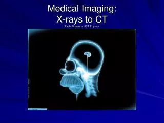
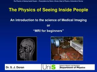
The Physics of Seeing Inside People A brief overview of medical imaging using MRI, PET, Ultrasound, X-ray CT and MEG Dr. S. J. Doran Department of Physics University of Surrey
Structure of the Talk • What is Medical Imaging? • Why use different methods of imaging? • Basic principles of imaging • Ultrasound • MRI • X-ray computed tomography (CT) • PET • MEG
What is Medical Imaging? Hi-tech scanner Images (preferably wacky colours) come out Patient goes in Medical imaging as seen by “Tomorrow’s World” !
Ultrasound Composite MRI + PET X-ray CT What is Medical Imaging? • The application of basic Physics to see inside the human body • Not one subject but many — lots of different techniques • Each one measures a different physical property of the sample.
Data source : Toshiba America Medical Systems Visualisation : Vitrea 2, Vital Images Data source : Mayo Clinic Why use different methods of imaging ? 1. Different methods reveal different features • Plane-film X-ray maps the total attenuation of X-rays along a path through the body, giving a projection image. Good for bone structure in accidents. • X-ray CT measures the X-ray attenuation coefficient of the body at each point. True 3-D images. • Ultrasound maps the reflectionand attenuation of sound.
Data source : FORENAP, Rouffach, France Data source : SMIS Ltd Data source : CSUA, Berkeley Why use different methods of imaging ? 1. Different methods reveal different features (cont.) • MRI maps the distribution and “environment” of water molecules in the body. • PET maps the distribution of radioactively labelled compounds. • MEG maps directly the magnetic fields generated by currents flowing in the brain.
Why use different methods of imaging ? 2. Accessibility and Portability A sliding scale MRI (most of the time!) … and almost everything else. Ultrasound MRI (sometimes!)
Why use different methods of imaging ? 3. Repeated exposure and safety The ALARP Principle - As Low As Reasonably Practiable • No medical imaging scan will be done unless the prescribing doctor is sure that it will not harm you. • Nevertheless, human biology is a complicated thing that none of us completely understands, so we try to keep all risks to an absolute minimum. • Different modalities of imaging use different types of radiation (e.g., MRI uses radio waves, PET uses gamma rays), which have different characteristics. At all times, we should pick the least invasive method.
Why use different methods of imaging ? 4. Patient acceptability • Different types of scan entail different degrees of patient discomfort. Some might require an injection of a contrast agent. The scanning environment is also important: Patient can be accompanied byPatient isolatedfriends or relatives. Open, comfortable environmentClaustrophobic tunnel Close contact with radiographerContact only via intercomduring scan procedure Patients can see images ofPatients do not know whatthemselves during the scan.is happening. UltrasoundMRI
Ultrasound X-ray CT MRI PET £500k - £1M £2M - £4M >£5M £20k - £100k Why use different methods of imaging ? 5. Cost Effectiveness • A good guiding principle in many walks of life is …Always pick the simplest solution for your problem. • In many cases the cheapest solution is the best.You do not need to give every pregnant mother in the country an MRI scan. • The capital cost of installation varies widely:
Reflector Emitted pulse 0 Transducer Lower amplitude reflected pulse Basic Principles of Diagnostic Ultrasound c c d • Based on ultrasound reflection and attenuation coefficients • Position calculated using equation d = ct/2
Use of Ultrasound in Obstetrics 5.5 Weeks 6 Weeks Data source : Joseph Woo
Use of Ultrasound in Obstetrics Bi-parietal diameter Length of femur Measurements of foetus in utero Data source : Joseph Woo
Use of Ultrasound in Obstetrics 18 Weeks 19 Weeks Data source : Joseph Woo
Use of Ultrasound in Obstetrics Duplex of flow in umbilical chord Duplex Measurements of blood flow on the foetus in utero Data source : Joseph Woo
Use of Ultrasound in Cardiology Standard real-time B-scan Duplex scan: colour Doppler super-imposed on real-time B-scan Diagnosis: Severe mitral regurgitation due to flail posterior MV leaflet. Underlying pathology: Mitral valve prolapse with ruptured chordae tendinae. Data source : Arizona Society of Echocardiography Image Library
Duplex Doppler Flow Data “Signed” velocity “Doppler Power” Flow pattern at the bifurcation of the carotid artery Data source : St. Paul’s Hospital, BC, Canada
Precession Spin Spin down N S Basic Principles of MRI • In a magnetic field, spinning nucleiprecess at the Larmor frequency. • The spin-up and spin-down nuclei have different energies. • Transitions between levels lead to emission of photons, which we can detect. Emitted photon Spin up Strong magnetic field
The Human Brain as seen by MRI Data sources : Left - The Whole-brain Atlas, K. A. Johnson and J. A. Becker, Harvard; Right - SMIS UK Ltd.
X-ray detector strip X-ray source Target 150 kV - + ~ Filament Basic Principles of X-ray CT • A standard tube produces X-rays with an energy of approximately 150 keV. • A fan-shaped beam passes through the patient and is detected to form a projection. • The source and detector are rotated to obtain a larger number of different projections. • Images are reconstructed using a technique called back projection and give a measure of the X-ray attenuation coefficient at every point.
X-ray CT Pictures of the Head and Abdomen Different densities of tissue give inter-mediate results Bone shows up bright Air is dark
g-ray detector + - Radioactive nucleus Basic Principles of Positron Emission Tomography (PET) • A radioactive isotope is injected and decays, emitting a b +-particle. • Within a short distance, the b +-particle bumps into an electron and the two annihilate, producing a pair of g -rays. • By detecting and reconstructing where the g -rays of come from, we can measure the location and concentration of radio-isotope.
A Typical PET Scanner Installation Cyclotron The PET scanner itself Radio-chemistry Lab Image Source: North Carolina Baptist Hospital/Bowman Gray School of Medicine; Children’s Hospital, University of Michigan
“Dead” areas of brainNo glucose metabolism Data source: Bowman Gray School of Medicine Two Typical PET Studies The highlighted region shows which part of the brain (the parietal lobe) was active during a visual stimulation task. Data source: CVVC, Psychology Dept., Durham Univ,. FDG Study of Patient with Stroke FDG “Brain Activation” Study
Principles of Magnetoencephalography (MEG) • The patient lies, in a magnetically screened room, next to a large number of very sensitive magnetic field detectors. • Electric currents within the brain create tiny magnetic fields — note the scale on the graph (fT, I.e., 10-15 times the magnetic field in an MRI scanner). • The complete set of measurements allows us to “solve” the “inverse problem” and find out what sort of current distribution created the magnetic fields. • We can then plot the result on, for example, an “anatomical” MRI scan. Data source: FORENAP, Rouffach, France
Image fusion — combining modalities Clinical Study: Lung Tumour Research study: functional imaging CT PET Fused Data source: Functional Imaging Laboratory, London Combined PET / MRI study
Typical uses of the different types of imaging (1) • X-ray imaging • Plane-film: bony trauma e.g., car crash • Fluoroscopy (real-time projection imaging, e.g., intra-operative) • CT: head scans involving fracture, bleeding scanning of other organs via addition of contrast agents • Ultrasound • Obstetrics • Flow and cardiac imaging • Applications requiring high portability of equipment • MRI • Method of choice for most soft tissue imaging • Cancer, stroke, multiple sclerosis, degenerative diseases • Functional brain mapping
Typical uses of the different types of imaging (2) • PET • Functional brain imaging using H217O or radio-labelled glucose (fluoro-deoxy glucose FDG). • Metabolic studies • Receptor studies • MEG • Epilepsy — location of seizure focus • Brain function • + other techniques not discussed in detail • SPECT — Single Photon Emission Computed Tomography • Optical imaging • Optical tomography
Conclusion • There are many different ways of imaging the human body. • The different methods tell us different things. • It is study of basic Physics (acoustics, magnetism, nuclear and particle physics) which has discovered the principles. • It is money — the human brain is a very valuable thing — which has led to the incredible developments that we see today.