Micro-Neuroscience
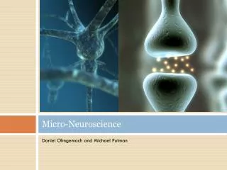
Micro-Neuroscience. Daniel Ohngemach and Michael Putman. Neurons and Glia. Cells of the Nervous System are divided into two types: Neurons are specialized to transmit information in the form of electrical signals. Glia serve more diverse roles: Myelination (Schwann Cells, Oligodendrocytes)

Micro-Neuroscience
E N D
Presentation Transcript
Micro-Neuroscience Daniel Ohngemach and Michael Putman
Neurons and Glia • Cells of the Nervous System are divided into two types: • Neurons are specialized to transmit information in the form of electrical signals. • Glia serve more diverse roles: • Myelination (Schwann Cells, Oligodendrocytes) • Nourishment (Astrocytes)
cell body Neuron Structure • Neurons exhibit two distinct features- the soma and neurites. • The soma, or cell body, is functionally equivalent to that of any other cell, but is differentiated to support neuronal activity. • There are two major classes of neurites- dendrites, which receive signals, and axons, which transmit signals.
The Axon • The axon is a long and thin extension of the cell membrane (and cytosol). • The composition of the axonal membrane and cytoplasm is different than the rest of the cell. • The Axon begins at the axon hillock, and can branch in multiple directions, ending at an axon terminal. • Important molecules are transported to the terminal via axoplasmic transport (along microtubules).
Biological Membranes • Cell membranes are composed mostly of phospholipids (bilayer). • The distinguishing feature of a membrane are its embedded proteins. • Many of these are integral membrane proteins: • Channels • Receptors • Connectors • Etc.
Channel Proteins • Ungated Ion channels allow for unregulated flow of ions down concentration gradients. • Gated channels regulate ion flow: • Voltage Gated- controlled by changes in membrane’s electrical potential. • Ligand gated- controlled by binding of a chemical. • The Sodium-Potassium Pump moves ions against gradients.
Resting Membrane Potential (RMP) • Various ion channels work in concord to generate an electrical potential across the membrane. • RMP in most human neurons: -65 mV. • The RMP is a result of Na+/K+ concentrations. • High concentration of Na+ outside the cell causes negatively charged ions to line up alone the inside of the membrane. • K+ concentration inside the cell is kept high by the Na+/K+ pump (also keeps Na+ concentration outside the cell high).
Action Potential (AP) • Nearby membrane is depolarized; voltage-gated Na+ channels open (“Threshold”). • Na+ ions flood the local axon. • Electrical Potential across membrane goes from RMP (-65 mV) to 32 mV. • Voltage-gated Na+ channels inactivate; voltage-gated K+ channels activate. K+ ions flow out of axon. • Membrane becomes hyperpolarized (<-65 mV)- high concentration of Na+ inside, K+ outside. • Na+/K+ pump restores RMP.
Synapses • Synapses fall into two major categories: • Electrical Synapses- created by physical connections between cells (Gap Junctions). • Chemical Synapses- open space (“synaptic cleft”) bridged by chemical neurotransmitters.
Synaptic Transmission • “Presynaptic Neuron” transmits an impulse to “Postsynaptic” Neuron • Presynaptic axon stores neurotransmitters in vesicles in the axon terminal. Neurotransmitter release is triggered by an influx of Ca++ ions, which cause neurotransmitter-containing vesicles to fuse with the membrane of the axon terminal. • Postsynaptic membrane contains receptors- proteins which bind neurotransmitters and effect a response. • Excess neurotransmitter release from the presynaptic axon is either recovered by the presynaptic cell or degraded enzymatically.
Receptors • Effect of a neurotransmitter depends on the action taken by the receptor. • Each receptor recognizes/binds a unique neurotransmitter (only 1!). • A receptor agonist (e.g. nicotine) mimics a neurotransmitter and a receptor antagonist (e.g. curare) inhibits the receptor’s behavior. • Two main types of receptor: • Ligand-gated ion channels allow ion flow when they bind neurotransmitters. • G-protein coupled receptors activate a second messenger, which may open ion channels or have some other effect.
Amines • Dopamine is involved in the brain’s reward system (reinforces good behaviors), and can lead to drug addiction. • Epinephrine release causes sympathetic activation (“fight or flight”). • Norepinephrine is thought to increase the brain’s responsiveness to stimuli (involved in attention, sleep, and a host of other systems). • Serotonin is the major neurotransmitter affecting mood and emotional behavior. Serotonin Dopamine Epinephrine Norepinephrine
Amino Acids • Glutamate is the major excitatory neurotransmitter of the brain- most commonly used to trigger Excitatory Postsynaptic Potentials (EPSPs). • Gamma-aminobutyric acid (GABA) is the major inhibitory neurotransmitter of the brain- most commonly used to trigger Inhibitory Postsynaptic Potentials (IPSPs).
Proteins • Peptide neurotransmitters are larger than others- this is thought to lead to slower release time. • Many are not used for direct neuron-neuron communication, but have other effects. • Substance P is a peptide neurotransmitter that is essential in the regulation of pain. Substance P
Other Neurotransmitters • Acetylcholine (Ach) is the main neurotransmitter at the neuromuscular junction (also exhibits effects in the brain). • Adenosine Triphosphate (ATP) is often found packaged in vesicles with other neurotransmitters, and is thought to enhance the effects of any given neurotransmitter. ATP
Diffuse Modulatory Systems • Small centers within the brain are responsible for overall concentration of given neurotransmitters. • Diffuse Modulatory neurons often synapse with over 100,000 other neurons. • As an example: Raphe nuclei, responsible for serotonin levels, are often found to be somewhat dysfunctional in many patients with clinical depression. Treatment often involves drugs known as Selective Serotonin Reuptake Inhibitors (SSRIs).
Motor Control • Muscle is innervated by alpha motor neurons. • Alpha motor neurons receive input directly from the spinal cord. • Each alpha motor neuron innervated several muscle fibers.
The Neuromuscular Junction • Excitation-Contraction Coupling: • ACh is released from alpha motor neuron, generating an action potential on the sarcolemma. • AP travels across sarcolemma and down transverse tubules. • AP activates voltage-regulated Ca++ channels in t-tubules. • T-tubule Ca++ channels are linked to large Ca++ release channels in the sarcoplasmic reticulum (SR), which stores large quantities of Ca++. When depolarized, release channels allow a large efflux of Ca++ into the cytosol of the muscle fibers.
Muscle Contraction • Major steps of contraction: • Ca++ binds to troponin, exposing binding sites on actin. • Myosin “heads” bind to the exposed sites on actin. • Binding causes myosin to “pivot.” • ATP hydrolysis provides energy to break myosin-actin bonds. • Cycle repeats- myosin “walks” down actin.
Memory • Memory is thought to be stored as neural circuitry- the path of impulse transmission. • Certain pathways are favored by increased conductance of a neuron when activated by a certain neurotransmitter. • Synaptic Plasticity: certain presynaptic-postsynaptic relationships are favored, mostly by an increased/decreased concentration of receptors in the postsynaptic membrane.
Memory: A Closer Look • One molecular mechanism of synaptic plasticity that is understood very well is AMPA/NMDA receptor augmentation. • Glutamate binds to postsynaptic receptors, leading to both ionotropic/metabotropic effects. • Second messengers can cause increased expression of receptor protein genes. Ions can activate protein kinases, activating receptors and causing them to be incorporated into the postsynaptic membrane.