Download
1 / 24
240 likes | 488 Vues
The Buccal Buckle: The Functional Morphology of Venom Spitting In Cobras Presented by: Qudirat Jamiu.

E N D
The Buccal Buckle: The Functional Morphology of Venom Spitting In CobrasPresented by: Qudirat Jamiu Young, B., Dunlap, K., Koenig, K., and Singer, M. “The buccal buckle: the functional morphology of venom spitting in cobras”The Journal of ExperimentalBiology Volume 207 (2004) Pages 3483-3494.
Background • Asiatic and African cobras independently evolved ability to expel venom as pressurized horizontal stream • Known as spitting • Form of active defensive behavior • Some cobras can spit venom as far as 3 meters • Pressure required • Most venomous snakes have to make direct physical contact with an object/organism to propel venom • Spitting cobras have the ability to expel venom without making direct physical contact
Have specialized exit orifice of the fang • Exit orifice is directed more craniad and has a more circular opening than the exit orifice of non-spitting cobras • This explains why venom is propelled forward, rather than downward. • Fangs surrounded by CT and epithelium • Attached to lateral and cranial surface of the maxilla • No smooth or skeletal muscle • Venom duct is also attached to lateral surface of maxilla
Jaw Anatomy • The upper jaw/ palato-maxillary arch of cobras consist of four bones • Pterygoid, ectopterygoid, palatine and maxilla • Four skeletal muscles contact the palato-maxillary • M. Protractor pterygoideus, M. levator pterygoideus, M. Retractor pterygoideus, and M. pterygoideus
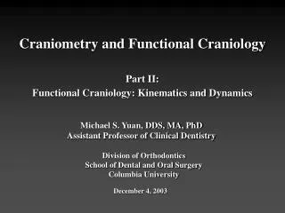
Illustration of the lateral view of the skull, and ventral view of the palato-maxillary arch, of Naja nigricollis. Muscle attachment sites are shown as solid colors (lateral surface) or broken colors (medial surface) for the following muscles: M. adductor mandibular externus superficialis (yellow); M. levator pterygoideus (purple); M. protractor pterygoideus (red); M. retractor pterygoideus (blue); M. pterygoideus (green). e, ectopterygoid; m, maxilla; pa, palatine; pf, prefrontal; pt, pterygoid.
Objective • To determine the functional bases of venom spitting • Muscle involvement • Bone Displacement
Materials and Methods • 5 Black-necked sitting cobras Naja nigricollis, 1 Red spitting cobra Naja pallida, 1 Indochinese spitiing cobra Naja siamensis, 4 Egyptian cobras Naja haje, and 3 Forest cobras Naja melanoleuca. • Venom expulsion measured using high speed videography and photography • Multiple spitting episode recorded using MotionScope 1000S at 500 frames s-1 • Recorded venom expulsions of spitting and non-spitting cobras
Sagital bisections of the heads of various species were examined to understand anatomy of spitting • Analysis of mechanical role of the M. protractor pterygoideus (PP) in spitting • Exposure of muscle • Applied bipolar stimulating probe to surface of mucsle • Electrical stimulations • Repeated stimulations with uniaxial strain guage
Venom pressure measurements • Fang removed • Polyethylene (PE) tubing placed over fang and attached to a pressure transducer • M. protractor pterygoideus (PP) and M. adductor mandibulae externus superficialis (AMES) exposed • AMES directly contacts venom gland, PP does not • Muscles were stimulated individually and simultaneously
Electromyography • (Instrument that produces an visual record of the electrical activity of a skeletal muscle by electrode inserted into the muscle or placed on the skin ) • Inserted into PP and AMES through incisions in head • Cobras were induced to spit by experimenter • Spit detector held in front of experimenters face • Striking sent signal to data system
Results • The high speed digital videography revealed that the spitting behavior had consistent patterns of motion • Four displacements occurred immediately prior to venom expulsion • Snout complex rotated in sagital plane elevating snout • Lateral displacement of caudal maxilla causing bulge/deformation of supralabial scales • Elevation of CT and epithelium surrounding fang to expose fang tip • Spit released with mouth slightly open at ~ 25o
Deformations of the palato-maxillary arch during spitting. (A) Naja nigricollis immediately prior to spitting; deformation of the supralabial scales (arrow) (B) a high-speed digital videograph recording of N. nigricollis spitting
The displacement of the palato-maxillary arch observed during spitting resembled the unilateral motions of the palato-maxillary arch associated with prey ingestion known as the ‘pterygoid walk’ • Displacements observed during spitting was never observed in non-spitting cobras, nor was it observed from spitting cobras engaged in any other behavior, including other forms of venom expulsion
Gross histological morphology of palato-maxillary arch of spitting and non-spitting cobras revealed minor differences except for the exit orifices • Contractions generated by electrical stimulation produced • displacements of palato maxillary arch • Protracted maxilla • Protracted palatine • Buckling of palato maxillary arch and maxilloectopterygoid joint
Ventral view of the palate of an Indochinese spitting cobra Naja siamensis before (A) and after (B) stimulation of the M. protractor pterygoideus. Both A and B are photos of the same side of the same animal Note the protraction and rotations of the palato-maxillary arch. f, fang; pa, palatine; pt, pterygoid.
12 electrical stimulations measured • Displacement of palatoperygoid and maxilloectopterygoidcan be seen by the signals obtained from the strain guage • Clear pattern of deformation observed prior to spits recorded Strain gauges placed on the palatal mucosa (A) or scales over the nasofrontal joint (B) of two separate spitting cobras Naja nigricollis. In both experiments contraction of the M. protractor pterygoideus, either artificially (A) or during spitting (B), resulted in deformations of the palato-maxillary arch evident in the strain gauge tracings.
Venom Pressure • Individual stimulations of PP and AMES produced clear patterns in pressure changes • AMES produced little pressure • PP produced displacement of palato-maxillary arch and tissue surrounding fang and produced pressure greater than that of the AMES • Two muscles stimulated together produced pressure greater than the sum of the individual muscle stimulations
Stimulation trials exposed to single twitch and train stimuli Venom pressure recorded from the fang tip of Naja nigricollis. Note that stimulation of the M. adductor mandibulae externus superficialis (AMES) simultaneously with the M. protractor pterygoideus (PP) produces greater venom pressure than when either is stimulated alone. This pattern holds whether the muscles are given twitch (A) or train (B) stimuli.
EMG activity • Electrical activity during spitting in the PP lasted 37 ms and in AMES 96ms • Mean spit duration 66ms • Palatal projections appeared 3 ms prior to onset of spitting
Conclusion • Analyses suggest that a two-component mechanism form the functional basis of venom spitting in cobras. • displacement of the palato-maxillary arch • increase in pressure caused by contraction of the M. adductor mandibulae externus superficialis • Spitting cobras utilize a pressure-balance system for venom expulsion; however, unlike all other venomous snakes where displacement of the fang sheath is passive, in spitting cobras the displacement is actively produced by the contraction of the M. protractor pterygoideus and the displacement of the palato-maxillary arch
Phylogeny of snakes remains uncertain, however functional convergence between Elapidae (rattlesnakes) and Viperidae (spitting cobras) has been suggested. • Venom delivery system of Elapidae and Viperidae • Nearly every aspect of the venom delivery system of elapids and viperids is morphologically distinct
It appears that rather than evolving a suite of morphological specializations for spitting, cobras have instead modified a motor action pattern employed for ingestion
Summary • Prior to spit discharge AMES involved in increasing venom pressure • AMES displaces the palato-maxillary arch • Contractions of the PP causes displacement of the palato-maxillary arch and fang sheath which also influences venom pressure • AMES and PP are activated simultaneously • This increases venom pressure within the venom gland, propelling stream of venom through the venom duct and out the fang