Download
1 / 84
910 likes | 1.63k Vues
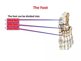
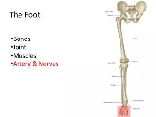
Chapter 18: The Foot. Arches of the Foot. Plantar Fascia. Joints and ligaments of the Foot. Muscle of the Foot and Lower Leg. Nerve Supply and Blood Supply. Functional Anatomy of the Foot and Biomechanics.

E N D
Functional Anatomy of the Foot and Biomechanics • ATC must realize that when considering foot, ankle, and leg injuries, that these segments are joined together to form a kinetic chain • Each movement of a segment, has an effect on proximal and distal segments • Lower-extremity chronic and overuse injuries have a number of biomechanical factors involved particularly when considering walking and running
Normal Gait • Two phases: • Stance or support phase which starts at initial heel strike and ends at toe-off • Swing or recovery which represents time from toe-off to heel strike
Foot serves as shock absorber at heel strike and adapts to uneven surface during stance • At push-off foot serves as rigid lever to provide propulsive force • Initial heel strike while running involves contact on lateral aspect of foot with subtalar joint in supination • 80% of distance runners follow heel strike pattern • Sprinters tend to be forefoot strikers • With initial contact there is obligatory external rotation of the tibia with subtalar supination • As loading occurs, foot and subtalar joint pronates and tibia internally rotates (transverse plane rotation at the knee)
Pronation allows for unlocking of midfoot and shock absorption • Also provides for even distribution of forces throughout the foot • Subtalar joint will remain in pronation for 55-85% of stance phase • occurring maximally as center of gravity passes over base of support • As foot moves to toe-off, foot supinates, causing midtarsal lock and lever formation in order to produce greater force
Subtalar Joint Pronation and Supination • Excessive or prolonged pronation or supination can contribute to overuse injuries • Subtalar joint allows foot to make stable contact with ground and get into weight bearing position • Excessive motion, compensates for structural deformity • Structural Deformities • Forefoot and rearfoot varus are usually associated with over-pronation • Forefoot valgus causes excess supination • May interfere with shock absorption
Excessive Prontation • Major cause of stress injuries due to overload of structures during extensive stance phase or into propulsive phase • Results in loose foot, allowing for more midfoot motion, compromising first ray and attachment of peroneus longus • Negative effect on pulley mechanism of cuboid relative to peroneal, decreasing stability of first ray • Causes more pressure on metatarsals and increases tibial rotation at knee • Will not allow foot to resupinate to provide rigid lever = less powerful and less efficient force produced
May also result in 2nd metatarsal stress fracture, plantar fascitis posterior tibialis tendinitis, Achilles tendinitis, tibial stress syndrome and media knee pain • Excessive Supination • Causes foot to remain rigid decreasing mobility of the calcaneocuboid joint and cuboid • Results in increased tension of peroneus longus and decreased mobility in first ray causing weight absorption on 1st and 5th metatarsals and inefficient ground reaction force absorption • Limits internal rotation and can lead to inversion sprains, tibial stress syndrome, peroneal tendinitis, IT-Band friction syndrome and trochanteric bursitis
Prevention of Foot Injuries • Highly vulnerable area to variety of injuries • Forces foot encounters can result in acute traumatic and overuse injuries • Injuries best prevented by selecting appropriate footwear, correcting biomechanical structural deficiencies through orthotics, and paying attention to hygiene
Selecting Appropriate Footwear • Numerous options available • Footwear should be appropriate for existing structural deformities (as evaluated by ATC) • For pronators a rigid shoe is recommended while supinators require more flexible footwear with increased cushioning • Basic form shoe is constructed on (last) dictates stability of shoe • Slip last shoe (moccasin style) is very flexible • Board last provides firm inflexible base • Combination last provides rearfoot stability and forefoot mobility
Shape of last may also determine selection • Straight-lasted vs. curve-lasted • Midsole design also set to control motion along medial aspect of foot • Heel counters are also used to control motion in the rearfoot • Other aspects of shoes that may impact foot include outsole contour and composition, lacing systems and forefoot wedges • Using Orthotics • Used to correct for biomechanical problems in the foot • Can be constructed of plastic, rubber, cork, or leather • Can be prefabricated or custom fitted
Foot Hygiene • By keeping toenails trimmed correctly, shaving down excessive calluses, keeping feet clean, wearing clean and correctly fitting socks and shoes and keeping feet as dry as possible to prevent development of athlete’s foot
Foot Assessment • History • Generic history questions • Questions specific to the foot • Location of pain - heel, foot, toes, arches? • Training surfaces or changes in footwear? • Changes in training, volume or type? • Does footwear increase discomfort? • Observations • Does athlete favor a foot, limp, or is unable to bear weight? • Does foot color change w/ weight bearing? • Is there pes planus/cavus? • How is foot alignment? • Structural deformities?
To assess structural deformities, subtalar neutral must be established • Draw line bisecting leg from start of Achilles tendon to distal end of calcaneus • Palpate the talus, inverting and everting foot so talus produces even pressure under index finger and thumb • Subtalar neutral
Once subtalar joint is neutral, mild dorsiflexion is applied to observe metatarsal head position relative to plantar surface of calcaneus • Degrees of forefoot and rearfoot valgus and varus can then be assessed • An equinus foot serves as a poor shock absorber • Forefoot is pronated relative to rearfoot when ankle is at 90 degrees of flexion • Similar to a plantar flexed first ray relative to the rearfoot
Shoe Wear Patterns • Over pronators tend to wear out shoe under 2nd metatarsal • Athletes often mistakenly perceive wear on the outside edge of the heel as being the result of over-pronation • Generally the result of the tibialis anterior causing foot inversion (while dorsiflexing) prior to heel strike to prevent foot from slapping ground • Wear on the lateral border of the shoe is a sign of excessive supination • Heel counter and forefoot should also be examined
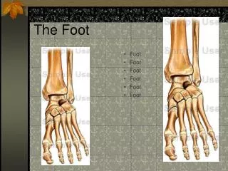
Medial calcaneus Calcaneal dome Medial malleolus Sustentaculum tali Talar head Navicular tubercle First cuneiform First metatarsal and metatarsophalangeal joint First phalanx Lateral calcaneus Lateral malleolus Sinus tarsi Peroneal tubercle Cuboid bone Styloid process Fifth metatarsal Fifth metatarsalphalangeal joint Fifth phalanx Palpation: Bony landmarks
Second, third and fourth metatarsals, metarsophalangeal joints, phalanges Third and fourth cuneiform Metatarsal heads Medial calcaneal tubercle Sesamoid bones Tibialis posterior Flexor hallucis longus Flexor digitorum longus Deltoid ligament Calcaneonavicular ligament Medial longitudinal arch Plantar fascia Transverse arch Palpation: Bony landmarks and soft tissue
Anterior talofibular ligament Calcaneofibular ligament Posterior talofibular ligament Peroneus longus tendon Peroneus brevis tendon Peroneus tertius Extensor hallucis longus Extensor digitorum longus tendon Extensor digitorum brevis tendon Tibialis anterior tendon Palpation: Soft tissue
Pulses • Must ensure proper circulation to foot • Can be assessed at posterior tibial and dorsalis pedis arteries • Dorsalis pedis pulse felt between extensor digitorum and hallucis longus tendons • Posterior tibial located behind medial malleolus along Achilles tendon
Special Tests • Movement • Extrinsic and intrinsic foot muscles should be assessed for pain, AROM, PROM, RROM • Tinel’s Sign • Tapping over posterior tibial nerve producing tingling distal to area • Numbness & paresthesia may indicate presence of tarsal tunnel syndrome
Morton’s Test • Transverse pressure applied to heads of metatarsals causing pain in forefoot • Positive sign may indicate neuroma or metatarsalgia
Neurological Assessment • Reflexes and cutaneous distribution of nerves must be tested • Skin sensation and alteration should be noted • Tendon reflexes (such as Achilles) should elicit a response when gently tapped • Sensation can be tested by running hands over all surfaces of foot and ankle
Recognition and Management of Specific Injuries • Foot problems are associated with improper footwear, poor hygiene, anatomical structural deviations or abnormal stresses • Sports place exceptional stress on feet • ATC’s must be aware of potential problems and be capable of identifying, ameliorating or preventing them
Injuries to the Tarsal Region • Fracture of the Talus • Etiology - • occurs either laterally from severe inversion/dorsiflexion force or medially from inversion/plantarflexion force with tibial external rotation • Sign & Symptoms - • history of repeated ankle trauma, pain with weight bearing, intermittent swelling, catching/snapping, talar dome tender upon palpation • Management • X-ray required for diagnosis, placed on weight bearing progression, rehab focuses on ROM and strengthening. If conservative management unsuccessful, surgery may be required (return to play in 6-8 weeks following surgery)
Fractures of the Calcaneus • Etiology • Occurs from jump or fall from height and often results in avulsion fractures anteriorly or posteriorly. • May present as posterior tibialis tendinitis • Sign and Symptoms • Immediate swelling, pain and inability to bear weight, minimal deformity unless comminuted fracture occurs • Management • RICE immediately, refer for X-ray for diagnosis • For non-displaced fracture, immobilization and early ROM exercises when pain and swelling subside
Calcaneal Stress Fracture • Etiology • Occurs due to repetitive trauma and is characterized by sudden onset in plantar-calcaneal area • Sign and Symptoms • Weight bearing (particularly at heel strike) causes pain, pain continues following exercise, • May require bone scan for diagnosis • Management • Conservative for 2-3 weeks, including rest AROM • Non-weight bearing cardio training should continue • As pain subsides, activity can be returned gradually
Apophysitis of the Calcaneus (Sever’s Disease) • Etiology • Traction injury at apophysis of calcaneus, where Achilles attaches • Sign and Symptoms • Pain occurs at posterior heel below Achilles attachment in children and adolescent athletes • Pain occurs during vigorous activity and ceases following activity • Management • Best treated with ice, rest, stretching and NSAID’s • Heel lift could also relieve some stress
Retrocalcaneal Bursitis (Pump Bump) • Etiology • Caused by inflammation of bursa beneath Achilles tendon • Result of pressure and rubbing of shoe heel counter of a shoe • Chronic condition that develops over time and may take extensive time to resolve, exostosis may also develop • Sign and Symptoms • Pain w/ palpation superior and anterior to Achilles insertion, swelling on both sides of the heel cord • Management • RICE and NSAID’s used as needed, ultrasound can reduce inflammation • Routine stretching of Achilles, heel lifts to reduce stress, donut pad to reduce pressure • Possibly invest in larger shoes with wider heel contours
Heel Contusion • Etiology • Caused by sudden starts, stops or changes of direction, irritation of fat pad • Pain often on the lateral aspect due to heel strike pattern • Sign and Symptoms • Severe pain in heel and is unable to withstand stress of weight bearing • Often warmth and redness over the tender area • Management • Reduce weight bearing for 24 hours, RICE and NSAID’s • Resume activity with heel cup or doughnut pad after pain has subsided (be sure to wear shock absorbent shoes
Cuboid Subluxation • Etiology • Pronation and trauma injury • Often confused with plantar fascitis • Primary reason for pain is stress on long peroneal muscle with foot in pronation • Sign and Symptoms • Displacement of cuboid causes pain along 4th and 5th metatarsals and over the cuboid • May refer pain to heel area and pain may increase following long periods of weight bearing • Management • Dramatic results may be obtained with jt. mobilization • Orthotic can be used maintain position of cuboid
Tarsal Tunnel Syndrome • Area behind medial malleolus forming tunnel with osseous floor and roof composed of flexor retinaculum • Etiology • Any condition that compromises tibialis posterior, flexor hallucis longus, flexor digitorum, tibial nerve, artery or vein • May result from previous fracture, tenosynovitis, acute trauma or excessive pronation • Sign and Symptoms • Pain and paresthesia along medial and plantar aspect of foot, motor weakness and atrophy may result • Increased pain at night with positive Tinel’s sign • Management • NSAID’s and anti-inflammatory modalities, orthotics and possibly surgery if condition is recurrent
Tarsometatarsal Fracture Dislocation (Lisfranc Injury) • Etiology • Occurs when foot hyperplantarflexed with foot already plantaflexed and rearfoot locked resulting in dorsal displacement of metatarsal bases • Sign and Symptoms • Pain and inability to bear weight, swelling and tenderness localized on dorsum of foot • Possible metatarsal fractures, sprains of 4th and 5th tarsometatarsal joints, may cause severe disruption of ligaments • Management • Key to treatment is recognition (refer to physician), realignment and maintaining stability • Generally requires open reduction with fixation • Complications include metatarsalgia, decreased metatarsophalangeal joint and long term disability
Injuries to Metatarsal Region • Pes Planus Foot (Flatfoot) • Etiology • Associated with excessive pronation, forefoot varus, wearing tight shoes (weakening supportive structures) being overweight, excessive exercise placing undo stress on arch • Sign and Symptoms • Pain, weakness or fatigue in medial longitudinal arch; calcaneal eversion, bulging navicular, flattening of medial longitudinal arch and dorsiflexion with lateral splaying of 1st metatarsal
Management • If not causing athlete pain or symptoms, nothing should be done to correct “problem” • If problems develop, orthotic should be constructed with medial wedge, taping of arch can also be used for additional support
Pes Cavus (High Arch Foot) • Etiology • Higher arch than normal; associated with excessive supination, accentuated high medial longitudinal arch • Sign and Symptoms • Poor shock absorption resulting in metatarsalgia, foot pain, clawed or hammer toes • Associated with forefoot valgus, shortening of Achilles and plantar fascia; heavy callus development on ball and heel of foot • Management • If asymptomatic, no attempt should be made to “correct” • Orthotics should be used if problems develop (lateral wedge) • Stretch Achilles and plantar fascia
Longitudinal Arch Strain • Etiology • Early season injury due to increased stress on arch • Flattening of foot during midsupport phase causing strain on arch (appear suddenly or develop slowly • Sign and Symptoms • Pain with running and jumping, usually below posterior tibialis tendon, accompanied by pain and swelling • May also be associated with sprained calcaneonavicular ligament and flexor hallucis longus strain • Management • Immediate care, RICE, reduction of weight bearing. • Weight bearing must be pain free • Arch taping may be used to allow pain free walking
Plantar Fasciitis • Common in athletes and nonathletes • Attributed to heel spurs, plantar fascia irritation, and bursitis • Catch all term used for pain in proximal arch and heel • Plantar fascia, dense, broad band of connective tissue attaching proximal and medially on the calcaneus and fans out over the plantar aspect of the foot • Works in maintaining stability of the foot and bracing the longitudinal arch
Etiology • Increased tension and stress on fascia (particularly during push off of running phase) • Change from rigid supportive footwear to flexible footwear • Poor running technique • Leg length discrepancy, excessive pronation, inflexible longitudinal arch, tight gastroc-soleus complex • Running on soft surfaces, shoes with poor support • Sign and Symptoms • Pain in anterior medial heel, along medial longitudinal arch • Increased pain in morning, loosens after first few steps • Increased pain with forefoot dorsiflexion