Download
1 / 37
380 likes | 617 Vues
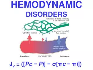
Infectious & Inflammatory Disorders. The lymphatic tissue in the neck accounts for 1/3 of all nodal tissue in the entire body Most cervical LN are located in the anterior triangle. Infectious lymphadenitis → extracapsular extension → neck space infection → frank neck abscess.

E N D
The lymphatic tissue in the neck accounts for 1/3 of all nodal tissue in the entire body • Most cervical LN are located in the anterior triangle
Infectious lymphadenitis → extracapsular extension → neck space infection → frank neck abscess
Acute bacterial lymphadenitis : Group A ß hemolytic strepococcus Staphylococcus aureus
Chronic form of lymphadenitis : Mycobacterium tuberculosis Atypical mycobacterium Cat-scratch disease Viral involvement Toxoplasmosis
Previous History • Age • Duration of symptom • Possible infectious contact • Animal exposure • Recent travel • Co-existing conditions
Physical inspection • Site • Size • Inflammatory characteristics : Tenderness Fluctuation Redness Warmness
Diagnostic Test • Needle aspiration • Excisional biopsy • Incision & drainage • Gram stain • Acid fast bacterial stain • Culture for aerobic & anaerobic bacteria • Viral ,fungal & unusual bacterial culture
Diagnostic test (cont.) • CBC • ESR • Serum Ig titer • TB skin test
Radiologic examination • Lateral neck X-ray • CXR • Axial CT scanning with IV contrast
Treatment Treatment for suppurative lymphadenitis is oral or IV broad-spectrum antibiotic with surgery reserved for refractory cases
Bacterial infection Penicillin First& second generation cephalosporin clindamycin
Viral infection Most common cause of cervical lymphadenitis RSV , parainfluenza , adenovirus ,HIV entrovirus, HSV , EBV Generalized LAP , Exanthems
Group A β hemolytic strep. & staph Unilateral cervical lymphadenitis 1-3 duration Level I & II Oral antibiotic
Cat-scratch disease last fall & winter 90% cat exposure ( Bartonella henselae ) Axillary LAP → Cervical LAP Skin lesion after 7-14 →1-2 weeks later tender lymphadenitis Can remain enlarged up to 4 month Needle aspiration may relieve acute pain Drainage should be avoided No treatment unless toxic symptom → Azithromycin
Mycobacterium species • The most common cause of chronic unilateral , suppurative cervical lymphadenitis • Positve tuberculin test will differentiated M. tuberculosis from atypical form . • Minimally tender , spontaneous rupture • Atypical form is rarely associated with pulmonary disease
Treatment • M. tuberculosis : six month rifampin, isoniazide ,pyrazinamide Atypical mycobacterium : Surgical excision with oral clarithromycin
Toxoplasmosis gondii • Infection via undercooked meat & unpasteurized milk • Immunocompetent → IM like viral infection • Immunocompromised → CNS infection • Oocytes in cat feces
Kawasaki disease • Fever • Rash • Mucositis • Nonpurulant conjunctivitis • Cervical lymphadenopathy • Common under 4 years • Toxin of S, aureous implicated as a possible etiology
Dental caries & gingivitis • Anaerobic species • Actinomycosis • Fungal • HSV
Fascial space infection • Submandibular space • Peritonsillar • Masticator space • Parotid space • Parapharyngeal space • retropharyngeal space
Pathophysiology of neck space infection • Pre-antibiotic era : tonsil & pharynx (70% ) , dentition ( 20% ) • Antibiotic era : Tonsil & pharynx (30%) , dental (30%) , cervical adenitis , trauma , IV drug abuser
Bacteriology • Mixed flora ( anaerobic abscesses predominating over aerobic abscesses) • Anaerobe : peptosterp. , Bacteriod , Anaerobic staph. • Aerobic : S. aureus , Strep. H. influ. E. coli , Klebsiella
Management of neck spaces infection • History of : • Recent dental work • Dental pain • IV drug abuse • URT infection • Recent surgery • Trauma
Management ( cont.) Physical examination : Inspection of dentition Palpation of the soft tissue of the H&N Vital sign
Radiologic study • Lateral neck film : may show increased prevertebral width : > 7 mm at C2 > 20 mm at C6 Presence of gas
Radiology ( cont.) • In patients who are initially managed with IV antibiotics , a CT scan should be used only when the patient is not progressing as expected . • Axial Ct with IV contrast
Wound cultures are not routinely obtained unless the patient is immunocompromised or the infection is unresponsive or life-threatening . 12-24 h of antibiotic therapy can differ cellulitis versus abcess .
Airway control is the first priority when the submandibular , parapharyngeal and retropharyngeal spaces are involved .
Severe trismus or Ludwig`s angina : Tracheostomy • Retropharyngeal abscess : Intubation in Trendelenburg position
Antibiotic • Clindamycin as a first – line coverage • Penicillin • Cefuroxime
Surgical drainage • Wide exposure , vascular identification • Drainage • Copious irrigation • Placement of drains ( 3-5 days )
Drainage (cont.) • Canine & sublingual space : intraorally • Buccal space : intra or extra oral • Masticator , submandibular ,submental : Extraoral parallel to the lower border of mandibule • Peritonsillar : aspiration & incision transorally
Drainage (cont.) • Parapharyngeal space : Transcervical • Retropharyngeal : transorally or transcervical • Parotid :standard parotidectomy incision
Necrotizing Fasciitis • Usually in trunk , extremity or perineum • Dental abscess , trauma , peritonsillar abscess , osteoradionecrosis , inset bite , burn , laceration , needle puncture • Predisposing factor : DM , peripheral vascular disease , cirrhosis , malignancy , alcoholism, immunosuppression
Progress over a few hours or a few days • Central zone of necrosis → tender purplish area → wide peripheral zone of erythema • Lack of frank purulence , thin gray exudate • Subcutaneous emphysema • Toxic state : hyperpyrexia , tachypnea , tachcardia , lethargy
Treatment • Correction of electrolyte imbalance , anemia , hypovolemia • Wide surgical debridement • Broad spectrum antibiotic • Aggressive bedside dressing • Frequent debridement under GA • Hyper baric oxygen