Download
1 / 16
160 likes | 339 Vues
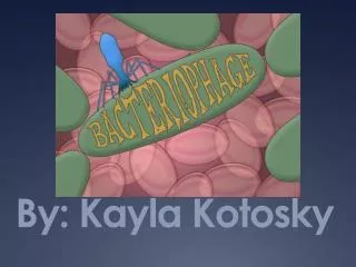
By: Kayla Kotosky. Intro to Bacteriophage. Virus that infects bacteria Reproduce by attaching to the outer surface of bacteria. Then inject viral genome into bacteria. Phage take over the cell’s replication to synthesize viral components. Lytic and Lysogenic Cycle.

E N D
Intro to Bacteriophage • Virus that infects bacteria • Reproduce by attaching to the outer surface of bacteria. Then inject viral genome into bacteria. Phage take over the cell’s replication to synthesize viral components.
Lytic and Lysogenic Cycle • In the lytic cycle virus particles replicate and bust out of the cell. • In the lysogenic cycle the virus does not immediately kill the infected cell. Instead it replicates with the host bacteria as it divides. • Virus particles are released by budding through the host cell, killing the cell.
What is Phage Therapy? • Bacterial infections can be treated with bacteriophages- culminating in their destruction. • Useful in medicine, veterinary science, dentistry, and even agriculture. • However, extensive testing still needs to be done before it becomes a first-line therapy.
Benefits of Phage Therapy • Skin grafting for wounds, burns, and skin cancer can be improved by phage therapy to lessen infections. • Bacteria causes food to spoil faster, phages have been studied to increase the freshness of food.
Methods • Collected soil samples from diverse locations. Our goal was to obtain a pure phage sample and sequence its DNA. • Most people had to switch to Hudson soil samples, because our samples didn’t obtain many, if any, viruses.
We took plaques that were provided for us (Hudson soil samples) and titrated them out. • We determined our phages titer, did a streak test, and a spot test to isolate more phages. • Our goal was to obtain a pure isolation of our plaques by obtaining a web plate. In order to get a pure isolation we didn’t want to have more than 1 plaque type on our plates.
Titrations • Most people did 3 or 4 purifications to make sure they had just one phage type. • Each time we picked 1 plaque and diluted it out to eventually obtain a pure sample, a web plate.
Spot and Streak Tests • For spot tests as our dilutions went down so did our titrations. • (10^0 would be the biggest spot and 10^-10 would have the smallest spot) • For streak tests we started with something very concentrated and we go to something with a lower concentration. So our samples became more dilute, had less phage.
What is MTL and HTL? • Then flooded our web plate from our purified titration = MTL • The MTL is a medium titer lysate, helped us find a specific plate we needed to harvest our HTL, which is a pure phage population.
Concentration of DNA • Now we wanted to isolate and purify our phages genomic DNA from our pure phage population. • Nuclease mix was added to our HTL’s to purity the sample and left to incubate. • After, we uncoated, isolated, and purified our DNA so we could calculate the concentration of our DNA.
Digestions • Digestions were then done with several different enzymes so we could get a variety of different cuts. • Different enzymes recognize and cut different sequences of DNA.
Group Digestion • 1. KB Ladder • 2. Undigested DNA • 3. BamHI • 4. ClaI • 5. EcoRI • 6. HaeIII • 7. HindIII
Gel Plate of my DNA Digestion • 1. KB Ladder • 2. Undigested DNA • 3. BamHI • 4. ClaI • 5. EcoRI • 6. HaeIII • 7. HindIII
2nd Gel Plate of my DNA Digestion • 1. KB Ladder • 2. Pst I • 3. Bcl I • 4. Nco I • 5. Eco RV
EM of my Phage • Head- 60nm • Tail- 100nm