The Human Cardiovascular System
This comprehensive overview of the human cardiovascular system includes essential details on its anatomy, principles, and functions. Dr. Larry M. Frolich outlines the two primary circuits: the pulmonary and systemic systems, and provides insights into heart structure, chambers, and valves. The text delves into the organization of veins, arteries, and capillaries, explaining how blood circulates to transport oxygen, nutrients, and hormones throughout the body. It also emphasizes the significance of cardiac function in maintaining overall health and discusses coronary blood supply.


The Human Cardiovascular System
E N D
Presentation Transcript
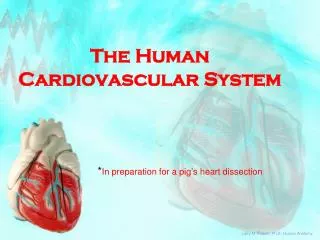
The HumanCardiovascular System *In preparation for a pig’s heart dissection Larry M. Frolich, Ph.D.,Human Anatomy
Cardiovascular/Circulatory System: • Principles • Structures • Two circuits • Pulmonary • Systemic • Heart Details • Other Details
CV System Function: • Circulate blood through entire body for • Transport of oxygen to cells • Transport of CO2 away from cells • Transport of nutrients (glucose) to cells • Movement of immune system components (cells, antibodies) • Transport of hormones
How? • Heart is pump • Diffusion in capillaries Veins = Return Arteries = Away
Overall Organization • Heart/Great Vessels--1 Route • Smaller arteries & veins --many routes -collateral circulation
Walls of Arteries and Veins • Tunica externa • Outermost layer • Strengthens, Anchors • Tunica media • Middle layer • Vaso-constriction/dilation • Tunica intima • Innermost layer • Minimize friction • Lumen
Capillaries • Microscopic: 1 cell thick • Network • Entire goal of C-V system is to get blood into capillaries where diffusion takes place
GREAT VESSELS • Aorta • IVC, SVC • Pulmonary Trunk • Pulmonary Veins
heartarteries arterioles veinsvenules capillaries 2 Circulatory Paths Systemic • Pulmonary
Heart Chambers and Valves Larry M. Frolich, Ph.D.,Human Anatomy
Right Heart Chambers: Pulmonary Circuit • Right Atrium (forms most of posterior of heart) • Receives O2-poor blood from body via IVC, SVC, Coronary sinus • Right Ventricle • Receives O2-poor blood from right atrium through tricuspid valve • Pumps blood to lungs via Pulmonary Semilunar Valve in pulmonary trunk • Septum
Left Heart Chambers: Systemic Circuit • Left Atrium • Receives O2-rich blood from 4 Pulmonary Veins • Left Ventricle (forms apex of heart) • Receives blood from Left Atrium via bicuspid valve • Pumps blood into aorta via Aortic Semilunar Valve to body
Heart Valves: Lub*-Dub** • *Tricuspid Valve: Right AV valve • 3 Cusps (flaps) made of endocardium and CT • Cusps anchored in Rt. Ventricle by Chordae Tendinae • Chordae Tendinae prevent inversion of cusps into atrium • Flow of blood pushes cusps open • When ventricle in diastole (relaxed), cusps hang limp in ventricle • Ventricular contraction increases pressure and forces cusps closed • *Bicuspid (Mitral) Valve: Left AV valve • 2 cusps anchored in Lft. Ventricle by chordae tendinae • Functions same as Rt. AV valve • **Semilunar valves: prevents backflow in large arteries • Pulmonary Semilunar Valve: Rt Ventricle and Pulmonary Trunk • Aortic Semilunar Valve: Left Ventricle and Aorta • 3 cusps: blood rushes past they’re flattened, as it settles they’re pushed down (valve closed)
Location of Heart in Thorax
Heart Wall • Epicardium (most superficial) • – Visceral pleura • Myocardium (middle layer) • Cardiac muscle • Contracts • Endocardium (inner) • Lines the heart
Blood supply to heart wall • Rt and Lft Coronary Arteries • Branch from Ascending Aorta • Have multiple branches along heart • Coronary Heart Disease • Cardiac Veins • Coronary Sinus (largest) • Many branches feed into sinus
http://www.rmgh.net/wiki/images/4/4b/Coronary_arteries_and_cardiac_veins.gifhttp://www.rmgh.net/wiki/images/4/4b/Coronary_arteries_and_cardiac_veins.gif