Thalamus & Limbic System
Thalamus & Limbic System. Objectives. By the end of the lecture, the student should be able to : Describe main functions of the thalamus Name and identify different nuclei of thalamus. Describe the connections and functions of thalamic nuclei.

Thalamus & Limbic System
E N D
Presentation Transcript
Thalamus & Limbic System
Objectives By the end of the lecture, the student should be able to: • Describe main functions of the thalamus • Name and identify different nuclei of thalamus. • Describe the connections and functions of thalamic nuclei. • Describe the effects of lesions of the thalamus. • Describe main functions of the limbic system. • Name and identify different parts of the limbic system. • Describe the effects of lesions of the limbic system.
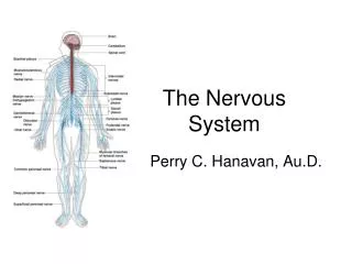
Thalamus • Part of diencephalon (other parts are hypothalamus, subthalamus and epithalamus) • A midline symmetrical structure, formed of 2 oval masses of grey matter • Is the largest nuclear mass • Situated between the cerebral cortex and brainstem Together with the hypothalamus, it forms the lateral wall of the 3rd ventricle T H 3rd ventricle
Thalamus It is the gateway to the cortex. It relays received information to the cerebral cortex from diverse brain regions. Axons from every sensory system (except olfaction) synapse in the thalamus as the last relay site before the information reaches the cerebral cortex. There are some thalamic nuclei that receive input from cerebellar nuclei, basal ganglia and limbic-related brain regions. Its function includes relaying sensory and motor signals to the cerebral cortex, along with the regulation of consciousness, sleep, and alertness.
RLV LLV S L M IC I 3rd V Thalamus has 4 surfaces • Superior • Inferior • Lateral • Medial: frequently connected to the thalamus of the opposite side by the interthalamic adhesion (massaintermedia)
Thalamus has 2 ends. *Anterior: Forms a projection called anterior tubercle which lies just behind the interventricular foramen. *Posterior: Forms a projection called Pulvinar which lies above the superior colliculus and the lateral & medial geniculate bodies. * * * Pulvinar. * Interventricular foramen.
Relations • Lateral: Posterior limb of the internal capsule (IC) • Medial: Together with hypothalamus, forms the lateral wall of the 3rd ventricle • Superior: Caudate nucleus (C) fornix (F) & lateral ventricle (LV) • Inferior: Hypothalamus (H) anteromedially & Subthalamus (ST) posterolaterally.
Internal Structure • Thalamus is mainly formed of grey matter divided by 2 sheets of white matter • External medullary lamina: • Covers the lateral surface separating the reticular nucleus from the rest of nuclei. • Consists of thalamocortical & corticothalamic fibers. • Internal medullary lamina: • Bundle of Y-shapedmyelinated (afferent & efferent) fibers. • Divides the thalamus into: anterior , medial, lateralnuclear groups. Extrnal Medullary lamina Each of these nuclear groups is subdivided into a number of named nuclei
Thalamic Nuclei • Anterior nuclear group: Anterior nucleus • Medial nuclear group: Largest nucleus is medial dorsal nucleus (MD) • Intralaminar nuclei: Lie within the internal medullary lamina • Midline nuclei: Lie deep to ependyma of 3rd ventricle
Thalamic Nuclei cont’d Lateral nuclear group is divided into Dorsal & Ventral tiers. • Dorsal tiercontains: • lateral dorsal n. (LD) • lateral posterior n. (LP) • pulvinar. • Ventral tiercontains • ventral anterior (VA) • ventral lateral (VL) • ventral posterior (VP) nuclei, divided into lateral & medial parts • medial & lateral geniculate bodies.
Functional Organization of Thalamic Nuclei • All thalamic nuclei EXCEPT reticular nucleus project to the ipsilateral cerebral cortex • Precise point to point projectionsexist between individual thalamic nuclei and restricted cortical zones. This type of nuclei are called ‘Specific nuclei’ All specific nuclei lie within the ventral tire of the lateral nuclear group. • All other nuclei are ‘Non-specific nuclei,
Classification of thalamic nuclei according to their projection • Simple sensory relay nuclei: receive well defined sensory impulses, and relay them to functionally distinct areas of the sensory cortex. • Ventral posterolateral nucleus (VPL). • Ventral posteromedial nucleus (VPM). • Lateral geniculate body (LGB). • Medial geniculate body (MGB). • They could be classified into 3 groups, each group contains 4 nuclei:
B) Circuit relay nuclei: receive impulses from different areas of CNS and relay them to specific areas in cerebral cortex. They include: Lateral ventral nucleus (projects to primary motor cortex). Anterior ventral nucleus (projects to premotor cortex). Anterior nucleus (projects to cingulategyrus). Part of dorsomedial nucleus. C) Associative nuclei: receive impulses from other thalamic nuclei and relay these impulses to the association areas of the cerebral cortex, They include: Part of dorsomedial nucleus. Pulvinar. Lateral dorsal nucleus. Lateral posterior nucleus.
The term "limbic" is from the Latin word Limbus, for "border" or "edge". • The limbic system is a set of evolutionarily primitive brain structures located on top of the brainstem and buried under the cortex • It separates the medial surface of the cerebral cortex from diencephalon Function of the limbic system It control a variety of functions including: • Emotions, emotional responses • Behaviour & mood (happy, cry, laugh, sad, fear, anger, aggression, depression) • Motivation. • Memory. • Visceral & Motor responses involved in sex, pleasure, hunger, and reproduction. • Olfaction.
The limbic system consists of a number of cortical& subcortical structures with complex and often looped connectionsthat all project to the hypothalamus. 1 8 4 6 10 3 7 9 2 5 1 The limbic system includes: Limbic lobe. Hippocampal formation. Septal area. Prefrontal area. Amygdala Anterior thalamic nuclei Hypothalamus (mammillary body) Fornix Olfactory system. Habenular nuclei
Limbic Lobe • C-shaped ring of grey matter on the medial side of each cerebral hemisphere, surrounding the corpus callosum. • It includes: • Subcallosal area • Cingulategyrus • Isthmus • Parahippocampalgyrus • Uncus. 2 1 3 5 4
Hippocampus • It is a seahorse shaped paired structure, one in each hemisphere. • Located in the inferomedial part of the temporal lobe. • Involved in formation, organization, storage and retrievalof memory
Its principal efferent projection is to the mammillary body via a C-shaped bundle of fibers called the Fornix. Fornix consists of: Fimbria Crus Body Column The fornix is an important component of PAPEZ CIRCUIT 3 2 4 1
Papez Circuit • 1937: Papez was the first to describe a relationship between limbic system components. • Papez’s circuit connects the parahippocampalgyrus, hippocampus, fornix, mamillary body, anterior thalamic nucelus and cingulategyrus. Since the initial description, connections to additional subcortical structures have been identified.
Hippocampal Formation Indusiumgersium Fornix • The hippocampal formation is a compound structure in the medial temporal lobe of the brain • It consists of: • Hippocampus • Dentate gyrus: Whichlies between hippocampus & Parahippocampal gyrus. • Subiculum(at the base of the hippocampus) • Entorhinal area (area 28) • Induseumgresium(grey matter on the upper surface of the corpus callosum). Entorhinal area Hippocampus Dentate gyrus
Amygdala • Almond shaped mass of nuclei, lies near the temporal pole, close to the tail of the caudte nucleus. Connections: • Input: from association areas of visual, auditory & somatosensory cortices. • Output: to hypothalamus & brainstem autonomic nuclei, to control the autonomic centers. Function: It is involved in emotional responses, fear, anger, hormonal secretions, and memory. Lesion: results in lack of emotional responses & docility
Septal Nuclei Site: Located anterior to the interventricular foramenbelow the rostrum of corpus callosum Main connections: • To hypothalamus throughmedial forebrain bundle. • Tohabenular nuclei throughstria medullaris thalami. Function: It provides critical interconnections and it is the pleasure zone. Septal area
Limbic Lobe Disorders • Korsakoff’spsychosis (Retrograde & anterogdrade amnesia) • Temporal lobe epilepsy: The hippocampus is a common focus site in epilepsy, and can be damaged through chronic seizures. • Alzheimer’s disease: Thehippocampus is one of the first brain areas to show damage in Alzheimer's disease • The hippocampus is sometimes damaged in diseases such as herpes encephalitis & Schizophrenia.