The Muscular System
230 likes | 340 Vues
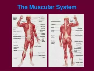
The human muscular system consists of three distinct types of muscle tissues: skeletal, smooth, and cardiac. Skeletal muscles, responsible for bone movement, are voluntary and work in pairs. Smooth muscles manage involuntary body functions and are found in organs like the stomach and intestines. Cardiac muscle, exclusive to the heart, ensures blood circulation. Each muscle type has unique structural characteristics and functions, including movement, posture maintenance, and regulating bodily functions. This overview provides insights into the complexities of muscle tissues.

The Muscular System
E N D
Presentation Transcript
Types of Muscle Tissue • All highly vascular for transport of oxygen, CO2, waste and sugars • Human body has three types: • Skeletal muscle: moves bones • Smooth muscle: body functions that you do not control consciously • Cardiac muscle: found in heart, pumps blood
Skeletal Muscle • Made of elongated cells called muscle fibers • Each fiber has many nuclei and striations • Striations: alternating light and dark bands • Skeletal muscles are voluntary muscles and work in pairs • Called antagonistic because it pulls against another type of muscle and vice versa
Function of Skeletal Muscles • Produce movement • Maintain posture and body position • Support soft tissue • Guard entrances and exits • Encircle openings to digestive and urinary tracts. Control swallowing, defecation and urination • Maintain body temperature
Smooth Muscle • Forms the walls of the stomach, intestines, blood vessels, and internal organs • Individual cells are spindle shaped with one nucleus • No striations • Surrounded by connective tissue • Involuntary muscle
Cardiac Muscle • Makes up the walls of the heart • Cells are cigar shaped • Intercalated disc: a double membrane separating adjacent cells in cardiac muscle fibers • Shares characteristics with both other types of muscle: • Is striated • Is involuntary • Has one nucleus
Muscle Structure • From the outside in… • Epimysium: tough, fibrous connective tissue that surrounds and separates fasciculi • Fascicle: bundle of muscle fibers • Perimysium: connective tissues that surrounds and separates fasciculi • Muscle fiber: muscle cell, large, long, cylindrical, multinucleated and made mostly of microfilaments • Sarcolema • Sarcoplasma • Sarcosomes • Sarcoplasmic reticulum • Endomysium
Muscle Structure • From the outside in continued… • Myofibrils – microfilaments (actin and myosin) that fill a muscle fiber • Actin: thinner protein made of two twisted filaments • Myosin: thicker protein, bigger molecule • These overlap, giving the striated appearance • Thin actin filaments are anchored to a structure called the Z line • Region from one Z line to the next is a sarcomere
Four Functions • Irritability • Responds to stimulation • Stimulation from nerves • Contractibility • Ability to shorten and move • Pulls on bones • Elasticity • Ability to resume original shape after contraction • Conductivity • Ability to carry an impulse • Causes contraction
Muscular Contraction • Sliding Filament Theory (Huxley) • Need stimulus from motor neuron for muscle to contract • Calcium ions are stored in sarcoplasmic reticulum • Very few calcium ions are found in the sarcoplasm of relaxed muscle • Muscle cells produce ATP, which sits on myosin in relaxed muscle • Motor End Plate: Terminal end of motor neuron that sits in a depression • Close to sarcolema but does not touch it (Space is called the synapse) • Comes from a motor neuron or a neuron that stimulates muscles to contract • Carries impulse to muscle or innervates muscle
Steps of Muscular Contraction • Action potential from the motor neuron moves down to terminal end and then calcium ions diffuse into the terminal • Calcium ions trigger the synaptic vesicles to release acetylcholine, a neurostransmitter • Acetylcholine diffuses across synaptic clef to a receptor site on the sarcolema • Acetylcholine cause new action potential to spread from nerve to muscle • Action potential spreads over the sarcolema until it finds an opening • It goes through the opening to the sarcoplasmic reticulum, where it triggers the release of calcium ions into the sarcoplasm • Calcium ions cause active sites on actin to be exposed • Myosin crossbridges can now link up with the active sites on actin • Actin moves inward, sarcomere shortens, muscle fibers contract and use a lot of ATP
Stopping Muscular Contraction • Acetylcholinesterase is released as muscle contracts and neutralizes the acetylcholine • No more action potential Is traveling to muscle • Calcium does not tighten so the crossbridges on mysoin release • Muscles relax
Moving Muscles • Muscles are attached to the outer layer of bone with a tough fibrous cord called a tendon • Origin: where muscle attaches to a relatively stationary position and anchors muscle • Insertion: where the muscle attaches to the moving bone • Action: result of muscle contraction; movement • Most muscles work in pairs; one contracts while one relaxes for smooth movement • Flexor: a muscle that bends a joint • Extensor: a muscle that straightens a joint


