NMR imaging
Learn about NMR imaging, a non-destructive and in-vivo imaging method with dynamic capabilities. Understand the principles behind NMR techniques like X-rays, ultrasound, and gamma rays, and how they are used for medical and veterinary purposes with excellent geometric resolution. Explore the fascinating world of NMR through detailed descriptions of processes, frequencies, and applications. Discover the high-contrast imaging capabilities of NMR without ionizing radiation exposure.

NMR imaging
E N D
Presentation Transcript
NMR imaging Mikael Jensen Associate professor Dept. Mathematics and Physics Royal Veterinary and Agricultural University April 2002
Why imaging • Non-destructive • Dynamic • In-vivo • or • Destructive • Static • Once in a lifetime
Veterinary use • Røntgen • Ultralyd • Termografi
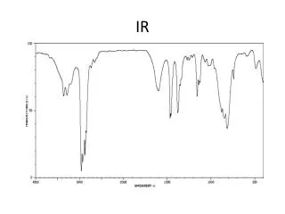
Penetrating radiation • X-rays (20-200 keV) • Gamma (80-511 keV) • Radiofreqency 63 MHz • Light(near infra-red) • Ultrasound
X-ray • Show differences in electron density • Small inherent contrast in soft tissue • Many photons means good S/N ratio • Good geometric resolution • Planar ( projections ) • Can be used for tomography ( CT-scan) • Uses ionizing radiation
Radio-isotopes (gamma radiation) • Function more than anatomy • Totally dependent on nature of tracer • Few photons, high contrast • Poor gometric resolution • Uses ionizing radiation
Ultrasound • Shows differences in sound velocity and density* • Tissue borderlines • Air-filled cavities creates shadows • Real-time • Cheap and safe • Interactive * Egentlig: Forskel i akustisk impedans !
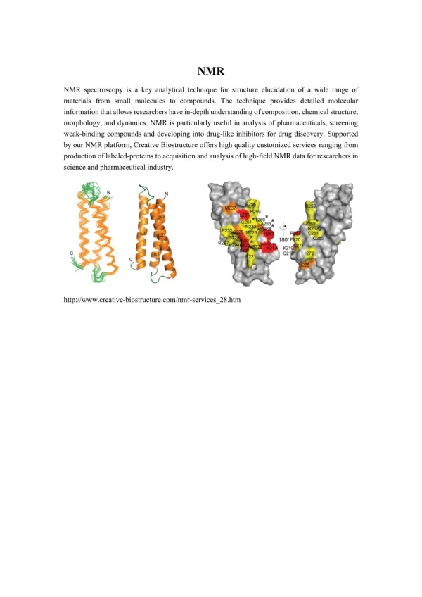
NMR – imaging (MRI) • No ionising radiation • Large inherent contrast in soft tissue • Can demonstrate both anatomy and function • Good geometrical resolution • Expensive • Restricted acces to patient during exam
Very good web introduction to MRI http://www.cis.rit.edu/htbooks/mri/inside.htm Go and read it !
NMR imaging Frequency= γ B For protons γ= 42 MHz / Tesla Wawelength at 1 Tesla ? Wawelength = c/f = 7 meter ! ?
External magnetic field necessary for NMR S z Bo y N x By convention we choose z axis along Bo
Nuclei Unpaired Protons Unpaired Neutrons Net Spin (MHz/T) 1H 1 0 1/2 42.58 2H 1 1 1 6.54 31P 0 1 1/2 17.25 23Na 0 1 3/2 11.27 14N 1 1 1 3.08 13C 0 1 1/2 10.71 19F 0 1 1/2 40.08 We need nuclei with magnetic moment for NMR Could be Hydrogen in water (protons)
Larmor condition E = h B E = h f f = B (Larmor condition) (proton)= 42 MHz/ Tesla When the energy of the photon matches the energy difference between the two spin states an absorption of energy occurs. In the NMR experiment, the frequency of the photon is in the radio frequency (RF) range. In NMR spectroscopy, is between 60 and 800 MHz for hydrogen nuclei. In clinical MRI, is typically between 15 and 80 MHz for hydrogen imaging.
Boltzman Statistics At room temperature, the number of spins in the lower energy level, N+, slightly outnumbers the number in the upper level, N-. Boltzmann statistics tells us that N-/N+ = e-E/kT. E is the energy difference between the spin states; k is Boltzmann's constant, 1.3805x10⁻²³ J/Kelvin; and T is the temperature in Kelvin. 1.000.000 1.000.001
T1 relaxation The time constant which describes how MZ returns to its equilibrium value is called the spin lattice relaxation time (T1). The equation governing this behavior as a function of the time t after its displacement is: Mz = Mo ( 1 - e-t/T1 )
T2 relaksation The time constant which describes the return to equilibrium of the transverse magnetization, MXY, is called the spin-spin relaxation time, T2. MXY =MXYo e-t/T2
Free induction decay (“FID”) Short RF pulse at Larmor frequency 90o Detected RF signal from nuclei
Fourier transform af FID F tid frekvens F-1 (sted)
NMR in organic chemistry CH3CH2OH Also known as Ethanol 1951 1991 Frequency alias ”chemical shift”
MRI imaging is ”broadband” • In chemical NMR typical resolution (linewidth) is 0.1 ppm • Chemical shifts are of the order of 1- 10 ppm • In imaging we have inhogeneous magnetic fields • In imaging we use frequncy to encode spatial position • Typical space coding 100 Hz/mm or 500 ppm/mm f f
Pulsewidth and flipangle 90o 180o 270o pw 2pw 3pw
Spin echo TE/2 TE/2 90o 180o
Gradient in magnetic field B = Bo + Gx x Bo = 1,5 T Gx = 25 mT /cm Frequency coding df/dx = Gx γ = 1 kHz /cm = 100 Hz/mm FFT time Gradient f
Imaging of one slice y x Gz z
Slice selected echo 90o 180o Only signal from slice Gz Normally chosen as z-direction
Read-out gradient 90o 180o Gz Gx
Phase encoding gradient 90o 180o Gz Gy Gx
Repeat this, and you got the image m data points 2D FFT m n n repetitions
Another way to do imaging Select one slice ! Do many experiments with different directions of readout gradient
Filtered back projection Radon transformation ( MRI, CT, PET, Spect ….) S.R. Deans, S. RoderickThe Radon Transform and Some of its Applications.Wilwy, New York1983
Slice selective MRI by back projection Many values Repeat for many angles Many values
Magnet B0 0.015 – 0.3 Tesla Resistive 0.5 – 3 Tesla Superconducting
Safety • Static magnetic field • No metal objects • Shielding • B < 3 Tesla • RF power deposition • Deposited power < 4 W / Kg • No hot spots • B < 3 Tesla (f < 130 MHz )
Lumbar spine MRI Prolaps Normal Malignancy ?
Liver Arrows point to multiple lesions in the liver demonstrating metastases.
Good image archives:NORTHEAST WISCONSIN MRI CENTERMR IMAGES http://www.newmri.com/humanbo2.htm RADIOLOGIC ANATOMY BROWSER™ http://rad.usuhs.mil/rad/iong/homepage.html
End of lecture Bloch Purcell Lauterbur