Download
1 / 33
370 likes | 693 Vues
rapid-prototyping of rapid-prototyping machines. Volume scanning Prof Phil Withers Manchester X-ray imaging Facility University of Manchester. From 3D object to 3D copy. 3D scanning. The first thing is to acquire a 3D virtual model of the item to be reproduced.

E N D
rapid-prototyping of rapid-prototyping machines Volume scanning Prof Phil Withers Manchester X-ray imaging Facility University of Manchester
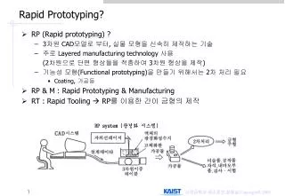
3D scanning The first thing is to acquire a 3D virtual model of the item to be reproduced. There are many different ways of scanning the object: • Contacting scanners • Non contact scanners
3D scanning (Contacting) Coordinate measurement machines • Very accurate • Can damage delicate samples • Very slow
3D scanning (non contact) Time-of-flight 3D laser scanner uses laser light to probe the subject. A laser is used to emit a pulse of light and the amount of time before the reflected light is seen by a detector is timed. • 10,000~100,000 points every second • operating over very long distances (km) • Good for buildings • mm accuracy Triangulation 3D laser scanners use laser to shine light on subject; exploits camera to look for the location of laser dot. • limited range (few meters) • accuracy is relatively high (~10microns). Hand-held laser scanners create a 3D image through triangulation Structured-light 3D scanners project structured pattern of light on subject and look at deformation of pattern on the subject. • Instead of scanning one point at a time, can scan entire field of view at once
Cheap 3D scanners • Makerbot 3d Scanner - nice little kit allowing you to use a normal consumer pico projector and webcam or other camera to make 3d scans of small objects • NextEngine– ~$3000, it does amazingly accurate scans, and is simple to use. • David 3D Scanner - uses a webcam and a handheld laser to allow you to scan in objects. The kit currently runs at about ~$1000. • Tgi3D PhotoScan– a software-based 3D scanning method using any digital camera to get your 3D model. You just take pictures of your object from different angles and use the software for 3D modeling with Google SketchUp.
Volume Scanning All the light based methods scan only the exterior envelope of the sample X-ray Computer Tomography (CT) • The great advantage of computer tomography is that not only do you get the external surface geometry you capture any internal features as well. • The principle is simple; namely to collect a series of radiographs (projections) acquired from different angles from which an image of the original 3D volume can be reconstructed using a computer algorithm • Range of resolutions from mm to tens of nanometers
Volume Scanning Magnetic resonance Imaging • Similar to x-rays but provides greater contrast for soft tissue • mm resolution
X-ray CT sources Laboratory x-rays Characteristic x-rays and broad spectrum
X-ray sources Broadly speaking X-ray penetration decreases with atomic number Broadly speaking X-ray penetration increases with energy to the 3rd power
X-ray Geometries • Spiral • Cone beam • Parallel beam
Contrast mechanisms (attenuation Contrast) • Let us consider the contrast in a 2D radiograph. • If the source is incoherent then features are recorded on the radiograph according to the attenuation of the x-rays along the travelled path • Consequently elements of high atomic number (e.g. Ca containing bones) attenuate more than those of low atomic number (e.g. C, H, O in soft tissue)
Contrast mechanisms (phase Contrast) • For low contrast features illuminated by coherent x-rays the phase change can be more significant than the attenuation change • By imaging at increasing sample to detector distances can increase the phase contrast. Phase contrast Attenuation contrast • Good for low contrast objects – e.g. fossils in amber, plastic objects, etc
CT tomography • Generally collect many projections (ideally ~ 1 for every 2 pixels across the detector) • i.e. for 2000 pixel detector use >1000 projections (radiographs) over 180° degrees • For complete solution need to capture whole width of sample in every image • Otherwise incomplete data (region of interest methods) Pixellated detector Spatial resolution usually >1pixel If object completely within detector Sample resolution ~width/No of pixels
CT Reconstruction • Once we have a set of projections we use a reconstruction algorithm to infer the 3D geometry of the object. • 99% of images are recovered using filtered back projection
CT Reconstruction (filtered back projection) (Parallel beam geometry) Each row of the sinogram (right) is the line of response (a pixel row on the detector) for a given projection (left)
CT Reconstruction (filtered back projection) By projecting the projections back at the angle for which they were collected you build up an image of the original slice – the more projections the better the image
CT Reconstruction Reconstruction of a metamorphic rock sample Attenuation of X-rays by the sample as a function of rotation Midsection of sample imaged with a planar fan beam Sinogram Horiz. – Detector channel Vert. – Rotation angle Brightness corresponds to extent of X-ray attenuation Each row of the sinogram is first convolved with a filter, and projected across the pixel matrix along the angle at which it was acquired http://serc.carleton.edu/research_education/geochemsheets/techniques/CT.html
CT tomography • The more projections the better the reconstructed image Restricted angles60 projections Only +/-60° (1 every 2°) 90 projections (1 every 2°) Restricted angles24 projections Only +/-60° (1 every 5°) 36 projections (1 every 5°)
Iterative reconstructions • Reconstruction of a 3D image from 2D image is an inverse problem. • Often not possible to exactly solve the inverse problem directly. • Iterative algorithms approach the correct solution using multiple iteration steps, allowing a better reconstruction at the cost of a higher computation time. • Large variety of algorithms, but each • starts with an assumed image, • computes projections from the image, • compares the original projection data and • updates the image based upon the difference between the calculated and the actual projections. Directreconstruction of real-time MRI of heart Iterativereconstruction
Iterative reconstructions There are typically five components to iterative image reconstruction algorithms: • A model of the object • A model of the measurement system/geometry • A statistical model of the noise. • A cost function that is to be minimized • An algorithm, usually iterative, for minimizing the cost function, including some initial estimate of the image and some stopping criterion for terminating the iterations. Comparison of FBP & iterative scheme for different total counts for image of liver. Note particular the streaking and noise appearance at low counts using FBP.
Iterative reconstructions Advantages: Lower dose (nosier data)
Iterative reconstructions Advantages:Phase segmentation (much better if wanting to extract a 3D solid model): use the number of phases as prior information (discrete tomography)
MESH PREDICT SCAN IMPROVE From tomograph to 3D model • Usually use thresholding to determine the boundaries of the geometry for the 3D solid model • Often the most difficult step is putting 3D model into CAD – can end up with models which are too complex and noisy and needs rationalising
Images to CAD • Many products for taking 3D models and importing into CAD, e.g. simpleware:
Preparing a scanned model for printing • The measured data alone, usually represented as a point cloud, lacks topological information • It must be processed and modeled into a more usable format such as a triangular-faced mesh, a set of NURBS surfaces, or a CAD model. • You need to clean up a model and fix it to be printable. There are a couple tools that are great for model cleaning.
Preparing a scanned model for printing Cleanup with Blender • Blender supports a TON of import and export formats. You'll want to export your final object as STL for printing though. • Remove duplicate vertices • Remove non-manifold points are points that just don't make sense in the real world. These can be hanging points, internal surfaces, holes, zero-thickness walls, etc. Clean STL file with netfabbwww.netfabb.com - alternative to blender easy automated tool- • netfabb studio basic is free • netfabbis an easy automated way to fix manifold and other mesh errors quickly.
Sending the image to a 3D printer Here is an example: • ReplicatorG, which is available at http://replicat.org/ and can be used to control printers such as makerbot • Once you have a 3D model, you need to run it through a slicer (e.g. skeinforge) to generate GCode, which is the file format that you send to the printer that tells it exactly what it needs to do in order to build your object. • Run the printer to create your 3D model layer by layer • Remove from substrate and finish off
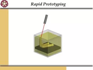
From 3D image to 3D object Subtractive Additive 3D machining 3D printing
Case study http://www.powerhousemuseum.com/collection/blog/index.php/2010/08/double-darwin-3d-scanning-and-rapid-prototyping-robot/
Case study Maker-bot : a DIY 3D printing system
Further reading http://wiki.makerbot.com/makerscanner http://www.david-laserscanner.com/ http://www.mxif.manchester.ac.uk/