Download
1 / 24
270 likes | 1.95k Vues
JOINTS. DR. ZAHID ALI KAIMKHANI M.D;M.Phil;Ph.D. ANATOMY DEPARTMENT COLLEGE OF MEDICINE KING SAUD UNIVERSITY. JOINTS. What do I need to know Explain the term “Joint”. Classify joints & describe each type with example. Describe the characteristics of synovial joints.

E N D
JOINTS DR. ZAHID ALI KAIMKHANI M.D;M.Phil;Ph.D ANATOMY DEPARTMENT COLLEGE OF MEDICINE KING SAUD UNIVERSITY Dr. Z Kaimkhani
JOINTS What do I need to know • Explain the term “Joint”. • Classify joints & describe each type with example. • Describe the characteristics of synovial joints. • Classify the synovial joint & describe each type with example. • List factors maintaining stability of joints. • Explain “Hilton’s law”.
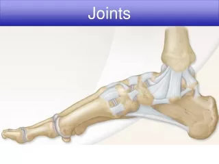
JOINTS- DEFINATION is the site where two or more than two bones meet together or union of two or more bones of the body. JOINT
JOINTS- CLASSIFICATION JOINTS are classified into: • Fibrous. • Cartilaginous. • Synovial. HOW?According to the tissues that lie between the bones.
JOINTS- CLASSIFICATION - FIBROUS Fibrous joints. The articulating surfaces are joined by fibrous tissue. Example: • SKULL SUTURES: No movement or negligible , temporaryas it ossify later in middle age). • INFERIOR TIBIOFIBULARJOINTS (SYNDESMOSIS): very Little movement, permanent joints. • GOMPHOSIS: Between teeth and their socket. Inferior
JOINTS- CLASSIFICATION -CARTILAGINOUS Cartilaginous joints. Union between bones is by cartilage. Two varieties: • Primary (Synchondrosis) • Secondary (Symphysis) • Primary Cartilaginous Joint : is one where bone and cartilage meet. • Immobile ,very strong • temporary joints, ossify later • Example • All epiphyses Between the Epiphysisand Diaphysis of a growing bone. • Junctions of ribs with their costal cartilage • Between the First Rib and the Sternum (1st sternocostal joint). Primary Cartilaginous
JOINTS- CLASSIFICATION -CARTILAGINOUS Cartilaginous joints. • Secondary Cartilaginous Joint :The bones are united by a plate of fibrocartilage. • Their articulating surfaces are covered by a thin plate of hyaline cartilage. • Little movement, permanent joints. • They are called Midline joints. • Examples: • Joints between the Vertebral Bodies (intervertebral discs). • Symphysis Pubis.
JOINTS- CLASSIFICATION –SYNOVIAL- CHARACTERISTIC Synovial joints. Characteristic features: • Fibrous capsule • Articular cartilage (Hyaline) • Joint cavity • Synovial membrane • Synovial fluid • Movements • Ligaments
JOINTS- CLASSIFICATION –SYNOVIAL- CHARACTERISTIC Synovial joints. Characteristic features: • The bones are joined by a fibrous capsule, which is attached to the margins of articular surfaces & enclosing the joint. • The articular surfaces are covered by a thin layer of hyaline cartilage (articular cartilage). • A joint cavity enclosed within the capsule. • A thin vascular synovial membrane lining the inner surface of the capsule. • A lubricating synovial fluid produced by synovial membrane in the joint cavity. The fluid minimizes the friction between the articular surfaces. • Movements. freely movable joints. • Ligaments . Reinforce the capsule externally or internally or both
JOINTS- CLASSIFICATION –SYNOVIAL-CLASSIFICATION Synovial joints. Classification Synovial joints can be classified according to: • The arrangement of the articular surfaces. • The types of movement that are possible
JOINTS- CLASSIFICATION –SYNOVIAL-CLASSIFICATION Synovial joints. Plane synovial joints • The articulating surfaces are flat. • The bones slide on one another, producing a gliding movement. Examples: • Sternoclavicular joint. • Acromioclavicular joint. • Intercarpal Joints.
JOINTS- CLASSIFICATION –SYNOVIAL-CLASSIFICATION Synovial joints. Axialsynovial joints Movements along different axes: • Transverse axis allows flexion & extension. • Longitudinal axis allows rotation. • Antero-posterior axis: allows abduction & adduction. Axial joints are divided into: • Uniaxial. • Biaxial. • Multi-axial (polyaxial).
JOINTS- CLASSIFICATION –SYNOVIAL-Axialsynovial joints Uniaxial. 1.Hinge joints: • Axis: Transverse. • Movements: Flexion & extension. Examples: • Elbow joint • Ankle joint. 2. Pivot joints : • Axis: longitudinal. • Movements: rotation. Examples: • Radio-ulnar • Atlantoaxialjoint. Elbow joint Atlantoaxial joint Radio-ulnar joint
JOINTS- CLASSIFICATION –SYNOVIAL-Axialsynovial joints Biaxial joints • Ellipsoid joints: • Saddle joints : • Ellipsoid joints: An elliptical convex fits into an elliptical concave articular surfaces. • Axis: Transverse & antero-posterior. • Movements: Flexion & extension, abduction & adduction BUT ROTATION IS IMPOSSIBLE. Example: • Wrist joint.
JOINTS- CLASSIFICATION –SYNOVIAL-Axialsynovial joints Saddle Biaxial joints (Conti) • Ellipsoid joints: • Saddle joints : 2. Saddlejoints : They resemble a saddle on a horse’s back. The articular surfaces are reciprocally concavoconvex. • Axis: longitudinal. • Movements: Flexion & extension, abduction & adduction and small range of ROTATION . Examples: • Carpometacarpal joint of the thumb.
JOINTS- CLASSIFICATION –SYNOVIAL-Axialsynovial joints Multi-axial Synovial joints Ball-and-Socket joints: • A ball –shaped head of a bone fits into a socket-like concavity of another. • Allows free movements • Movements: Flexion & extension, Abduction & adduction and Medial and lateral rotation. Examples: • Shoulder joint. • Hip Joint. Socket Ball
JOINTS - STABILITY Factors Affecting Stability of Synovial Joints • The shape of articular surfaces • Ligaments • Tone of muscles around the joint. • Atmospheric pressure
JOINTS - STABILITY Factors Affecting Stability of Synovial Joints • The shape of articular surfaces • The ball and socket shape of the Hip joint is a good examples of the importance of the shape of the bone, to maintain joint stability. • The shape of the bones forming the Knee joint has nothing to do for stability.
JOINTS - STABILITY Factors Affecting Stability of Synovial Joints 2. Strength of the ligaments: • They prevent excessive movement in a joint.
JOINTS - STABILITY Factors Affecting Stability of Synovial Joints 2. Tone of the surrounding muscles. • In most joints, it is the major factor controlling stability. • The short muscles around the shoulder joint (rotator cuff) keeps the head of the humerus in the shallow glenoid cavity.
JOINTS - NERVE SUPPLY NERVE SUPPLY OF JOINTS The capsule and ligaments receive an abundant sensory nerve supply. • Hilton’s Law “A sensory nerve supplying a joint also supplies the muscles moving that joint and the skin overlying the insertions of these muscles.”
JOINTS - SUMMARY Joint is the site where two or more bones come together, whether movement occurs (or not) between them. Joints are classified according to the tissues that lie between the bones into 3 types: fibrous, cartilaginous & synovial. Synovial joints are freely movable & characterized by the presence of: • Fibrous capsule, • Articular cartilage, • Ligaments • Synovial membrane & • Joint cavity containing synovial fluid. • Synovial joints are classified according to the range of movement into: plane and axial. • Axial are divided according to the number of axes of movements into: uniaxial, biaxial & polyaxial or multiaxial. • Stability of synovial joints depends on: shape of articular surfaces, ligaments & muscle tone. • Joints have same nerve supply as muscles moving them (Hilton’s law).
JOINTS - SELF ASSESSMENT Self Study Intra-articular fibrocartilages? Fatty pads? QUESTION “I” Which of the following is a pivot synovial joint? • Shoulder. • Ankle. • Sternoclavicular. • Atlantoaxial joint QUESTION “2” Which of the following is a Primary cartilaginous joint? • Hip. • Elbow. • Inferior radioulnar. • Symphysis pubis. • QUESTION “3” • Which of the following stabilizing factor is the most important in shoulder joint? • Articular surface. • Capsule • Muscles • Ligaments. • QUESTION “4” • The normal level synovial fluid in KNEE joint is? • 0.5 ml. • 5.0 ml. • 10 ml • 50 ml
THANK YOU Dr. Z Kaimkhani