Download
1 / 12
120 likes | 144 Vues
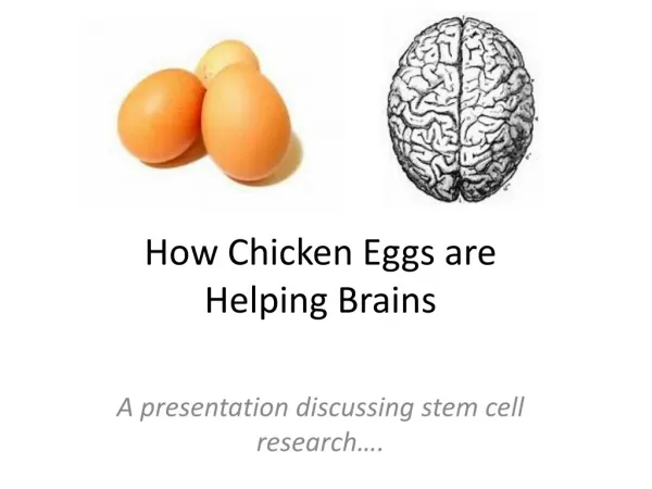
This article explains the fertilization and early development of chicken eggs, including the formation of the zygote, ectoderm, endoderm, and mesoderm layers. It also discusses the influence of temperature on embryonic development and the functions of the extra-embryonic membranes.

E N D
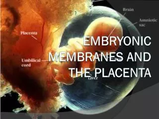
Extra Embryonic Membranesof Chicken Eggs Yassar Abbas Assistant Professor, Poultry Production CVAS,Jhang
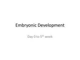
Fertilization and Early development • Fert. of the germinal disc by the sperm takes place in the infundibulum • about 15 minutes after its holding follicle has released the yolk. • Cell division to create the new embryo starts about 5 hrs after fertilization and continues
the egg passes along the oviduct and is eventually laid. • It is said that the hen’s egg takes 21 days of favorable incubation conditions for the chicken to develop and hatch. • However, this development takes 22 days – one day in the oviduct and 21 days in the incubator or nest
The zygote • When the sperm cell (with half the required chromosomes) fertilises the female egg cell (with the other half of the required chromosomes) • it forms the zygote – a single cell with the correct number of chromosomes. About 5hrs after fertilization the zygote enters the isthmus and it is here that the new embryo starts to develop by simple cell division. • By the time the egg leaves the isthmus, the zygote, now called the blastoderm or embryo, comprises 8 cells and after 4 hrs in the uterus it has grown to 256 cells.
Formation of ectoderm, endoderm and mesoderm • Initially the dividing cells form one layer over the yolk, but as cell division continues two layers are formed. These are called the • Ectoderm (uppermost) and the • Endoderm (underneath) layers. At about this stage the central cells of the blastoderm separate from their contact with the yolk to form a cavity. It is in this cavity that subsequent embryo development occurs. Soon after the formation of the ectoderm and endoderm, • a third layer of cells called the Mesoderm or middle layer is formed.
3 layers • The Ectoderm produces the nervous system, parts of the eyes, the feathers, beak, claws and skin. • The Endoderm produces the respiratory system, the digestive system and secretary organs • The Mesoderm produces the skeleton,muscles, circulatory system, reproductive organs and excretory system
Physiological zero • As reptilian ancestors • the influence of ambient temperature during the post laying period on embryonic development. • When the temperature of the egg is below 20°C, • the embryo becomes dormant and most development stops. • When the temperature rises above about 20°C, embryonic activity starts again. • This temperature of about 20°C when embryonic activity starts or stops is often referred to as • Physiological zero.
Extra-embryonic membranes • Yolk sac: This sac envelops the yolk, produces an enzyme that changes the yolk material into a form that can be used as a food source by the developing embryo. Any remaining, unused yolk material in the yolk sac when the chicken hatches from the egg is drawn into the abdomen for use by the chick for the first two to three days after hatching while the chick learns what to eat/drink and where to find it.
Amnion: • The amnion forms a sac • that is filled with fluid in which the embryo floats. • In this way it provides a • shock-absorbing environment in which the • fragile embryo can develop without harm from normal day to day knocks
Allantois: • The allantois develops an • extensive circulatory system connected to that of the embryo and driven by the new embryonic heart • When the allantois is fully developed it completely surrounds the embryo. • This membrane has a number of functions:
Respiratory • The developing embryo uses oxygen and produces carbon dioxide i.e. it has respiration. It is unable to carry out this function for itself and hence the allantois oxygenates the blood and eliminates carbon dioxide. Digestive • it provides the means for the embryo to access the albumen and the calcium of the shell
Chorion: • Excretory – • It removes the wastes that result from the embryo’s metabolism and deposits it in the allantoic cavity. • The chorion fuses the inner shell membrane to the allantois and helps that membrane to carry out its functions.