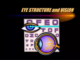
The Eye and Vision
690 likes | 905 Vues
Dive into the intricacies of the eye's structures and functions, from the cornea to the retina. Discover how light, shapes, and colors are processed to create vision, and learn about accessory structures like the eyelids, conjunctiva, and more.

The Eye and Vision
E N D
Presentation Transcript
The Eye and Vision Unit 2 Communication, 2.4 Communication with the Outside World
The Eye • The eye is a complex organ composed of many parts. • Good vision depends on the way in which those parts work together. • What we see is actually light that is differentiated into shapes and colors.
Our eye is about the size of a ping pong ball but slightly asymmetrical. • Has a length of 24 – 25 mm, and a diameter of about 24 mm. • Size Chart http://www.noadi.net/EyeSizes.html
Accessory Structures of the Eye • Eyebrows • Shade the eyes • Prevents liquids from the forehead going into the eye • Eyelids • Protects the eye • Has an inner layer known as the conjunctiva – covers the eye and secretes mucous • Helps spread liquids across the eyeball • Oils, mucus, saline
Accessory Structures Continued • Eyelashes • Responsible for sensing an object and cause use to blink in response – protective • Extrinsic Muscles • Allows the eye to move
Accessory Structures Continued • Lacrimal Apparatus • Lacrimal gland • Secretes tears –diluted salt solution, antibacterial enzyme • Lacrimal canal • Collects tears • Lacrimal sac • Collects the tears and passes them into the nasal cavity
Conjunctiva • A thin, transparent membrane that’s involved in protecting your eyes. • Lines the insides of the eyelids. • Has tiny blood vessels that supply it with nutrients.
Cornea • Transparent, domed-shaped structure at the front of the eye. • Due to lack of blood vessels • It allows light to pass through it before entering the eye. • It helps refract light (bending of light) when it enters the eye. • It is continuous with the sclera. • It heals quickly if damaged • Laser
Sclera • The “White” of the eye. • Serves as the eyes’ protective coat. • Muscles attach to it to move the eye. • It extends to the back of the eye to the optic nerve.
Iris and Pupil • The iris is a membrane in the eye, responsible for controlling the diameter and size of the pupil and the amount of light reaching the retina. • "Eye color" is the color of the iris. • In response to the amount of light entering the eye, muscles attached to the iris expand or contract the aperture at the center of the iris, known as the pupil. • The larger the pupil, the more light can enter.
The Lens • A transparent, biconvex structure in the eye. • Along with the cornea, helps to refract light to be focused on the retina. • By changing its shape, it functions to change the focal distance of the eye • Allowing for focus on objects at various distances = production of a sharp images on the retina.
Adjustments of the lens are made by the cillary body. • Contains suspensory ligaments (Zonule of Zinn) that hold the lens in place • The ligaments are moved by cillary muscles which cause the lens to change shape.
Choriod • A membrane found between the sclera and the retina, lines the back of the eye. • It contains many blood vessels that supply oxygen and nutrients to the retina, and is highly pigmented to help absorb light and prevent scattering. • The iris is the visible part of the choriod which gives the eye its color.
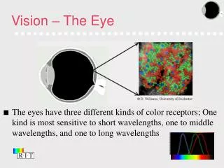
Retina • Lines the inside of the back part of the eye, and is the light-sensitive part of the eye. • It contains millions of cells known as photoreceptors, and each photoreceptor is linked to a nerve fiber. • Light signals are converted into nueral signals. • Rods and Cons
Blind Spot • In the retina • Does not contain photoreceptors • It is in this region that the optic nerves come together and exit the eye on their way to the brain. • An image that falls in this spot will not be seen. • Hopefully, the other eye can see the object.
The macula • Located in the center of the retina. • Light narrows to this point of the retina. • Gives you the sharp vision that is needed to do things such as read. • It is yellow in color.
The fovea • The absolute center of the macula. • Provides for the most clear vision. • There are NO rods...only cones at the fovea. • The cones are also packed closer together in the fovea than in the rest of the retina. • Blood vessels and nerve fibers go around the fovea so light has a direct path to the photoreceptors.
Aqueous Humor • Fills the space between the cornea and iris. • It is continually produced by the ciliary body, the part of the eye that lies just behind the iris. • This fluid nourishes the cornea and the lens and gives the front of the eye its form and shape.
Vitreous Humor • It is the clear gel that fills the space between the lens and the retina. • It is “stuck” to the retina. • It is transparent and colorless. • Unlike the fluid in the frontal parts of the eye (aqueous humour) which is continuously replenished, the gel in the vitreous chamber is stagnant. • It can detach from the retina causing floaters. • Purpose – to give the eye its shape
So….How do we See?How do the structures work? • Light rays go into the cornea • The cornea bends/refracts the light through the pupil • The pupil adjusts to the amount of light • Bright light = pupil constricts • Dark = pupil becomes large
The rays pass through the lens • Changes shape depending on distance of the object you are looking at • http://library.thinkquest.org/C005949/anatomy/lensciliary.htm • Focuses the light on the retina • Rods and Cons • Macula • Fovea • The image is upside down at this point • The cells of the retina sends electrical images to the optical nerve • Message travels to the brain and is deciphered into an image – right side up.
Photoreceptors of the Eye • In the Retina
Photoreceptors are not distributed evenly throughout the retina. • Most cones lie in the fovea, whereas peripheral vision is dominated by rods. • Overall, rods greatly outnumber cones. • rods (black and white vision, very sensitive to low light) • Better at detecting motion • cones (color vision, not so sensitive) • Used for visual resolution • Fovea – green and red cones • Blue cones most are location outside of the fovea. Add the underlined sections to your notes.
Rods • The light-sensitive pigment in rods is rhodopsin, • breaks down into a protein, opsin, and retinal (from vitamin A) in the presence of light. • Decomposition of rhodopsin activates an enzyme that initiates changes in the rod cell membrane, generating a nerve impulse. • Nerve impulses travel away from the retina and are interpreted as vision.
Cones • The light-sensitive pigments in cones are also proteins; there are three sets of cones, each containing a different visual pigment. • The wavelength of light determines the color perceived from it; each of the three pigments is sensitive to different wavelengths of light. • The color perceived depends upon which sets of cones the light stimulates: if all three sets are stimulated, the color is white; if none are stimulated, the color is black.
The Layers • All vertebrate retinas are composed of three layers of nerve cell bodies and two layers of synapses. • The outer nuclear layer contains cell bodies of the rods and cones • The inner nuclear layer contains cell bodies of the bipolar, horizontal and amacrine cells • The ganglion cell layer contains cell bodies of ganglion cells and displaced amacrine cells.
R = Rod • C = Cone • H = Horizontal cells • Lateral connection • between rods and • cones • B = Bipolar Neurons • A = Amacrine cells • Lateral connection bipolar and ganglion cells • Interneuron cells – connecting neurons • G = Ganglion Cells • Neuron cell that connects information from other neurons and transmits it to optic nerve and brain.
Occipital Lobe • The occipital lobe is where our images are processed.
Color Vision - review • The retina of the eye possesses two special types of nerve cells known as photoreceptors. • Rods function in dim light and perceive shades of gray. • Cones function in bright light and provide sharp, colorful images. • Impulses from rods and cones pass through nerve cells to the optic nerve.
Color Vision - Review • There are three different types of cones: • Red cones • Blue cones • Green cones • Each type of cone is sensitive to a different range of wavelengths of light. • Different types of cones function together to interpret colors other than blue, red, and green. • If any of the cones malfunction, color deficiency or color blindness occurs.
Depth Perception • Depth perception is the ability to judge the relative distances between objects in three dimensions. • With one eye, the field of vision appears two-dimensional. • With two eyes, the eyes see and the brain processes different views of the same object.
Accommodation • Two parts of the eye – the cornea and the lens – focus light on the retina. • The cornea does most of the work, but it cannot change shape. Fine adjustments are carried out by the lens. • Accommodation is the combination of reflex actions by which the lens of the eye changes to keep the focal length, the distance between the center of the lens and its focal point, constant. • Ciliary muscles in the eye assist adjustment of the lens.
Astigmatism • Astigmatism is a condition in which the cornea or the lens is irregularly shaped. • This shape change causes incoming light rays to refract and converge improperly. • The light rays do not focus at a specific point on the retina, resulting in a blurry or distorted image. • Astigmatism may be corrected with eyeglasses, contact lenses, or refractive surgery.
Blind Spot • The optic nerve exits the eye at the retina on its way to the brain. • Since this area of the retina does not have receptors that respond to light, it is referred to as the blind spot. An image that falls on this area can not be seen. • Normally people do not recognize the blind spot because the eyes are always on the move and the brain ignores this “hole” in visual input. • http://serviceworksconsulting.com/blind_spot/body_blind_spot.html
Peripheral Vision • Peripheral vision is the ability to see things that fall outside of the direct line of vision. • Due to their proximity to the edge of the retina, rods are responsible for this aspect of vision. • Peripheral vision is better for detecting movement than for processing sharp images, and is most often stronger in the dark.
Optical Illusions • Optical illusions are visual tricks that actually take place in the brain rather than the eye. • The visual cortex of the brain deciphers images sent from the eye, however surrounding objects, intense colors, distortions of expected patterns, and preconceptions can cause the mind to “see” and interpret an image differently. • http://www.scientificpsychic.com/graphics/
Afterimages • Afterimages are optical illusions that occur when looking away after staring intently at a fixed image or color. • The constant light stimulating the retina causes the cones in that area to become fatigued. • After looking away from the image, the less-stimulated cones, which are not fatigued, still function. • The resulting image lasts briefly and because it comes only from the less-fatigued cones, is perceived as a negative image.