Download
1 / 10
100 likes | 144 Vues
Dive into the intricate world of the human eye and vision system, exploring the layers of the retina, photoreceptors, and color perception. Learn about the fascinating mechanisms of cones and rods, adaptation to light levels, and the distribution of photoreceptors across the retina. Discover how the network of nerve cells in the retina processes visual information through lateral inhibition, creating optical illusions like Mach Bands and the Hermann Grid. Gain insights into human vision and color response.

E N D
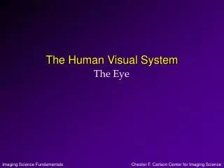
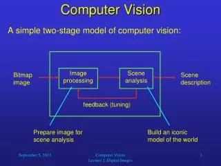
Plexiform Layer • The retina is made of three layers: • Plexiform layer is a network of nerves which carry the signals from the photo receptors. • Photo receptors. • Choroid provides nourishment to the receptors, as well as absorb any light that didn’t get absorbed by the photo receptors, like a antihalation backing in film. Fovea Photo receptors Light Plexiform Layer Choroid Optic Nerve
Rods and Cones Synaptic endings • Highly sensitive to low light level or scotopic conditions. • Black and white. • Dispersed in the periphery of the retina. Cell nucleus Inner segments Outer segments Rod Cone • Sensitive to high light level or photopic conditions. • Three types of cones responsible for color vision. • Concentrated in the fovea.
Adaptation • Why can’t you see immediately after you enter a movie theater from daylight? • The threshold of detection changes with overall light level. • The switch is quite gradual, until the sensitivities of cones and rods cross over at about 7 minutes in the dark. Photopic (cones) Scotopic (rods) Threshold of detection (log scale) 0 5 10 15 20 25 30 Time in dark (minutes)
80º 80 º 60 º 60 º 40 º 40 º 20 º 20 º 0 º Distribution of Photoreceptors Visual Axis • Cones are concentrated in the fovea. • Rods predominate the periphery. • There is a blind spot where there are no photoreceptors, at the point where the nerves exit the eye (optic nerve). Temporal Nasal Blind spot 160 140 Rods 120 Number of receptors per mm2 100 80 60 40 Cones 20 40 º 60 º 20 º 40 º 20 º 0 º 60 º 80 º Angle
S I L Relative response 400 460 490 500 530 600 650 700 Wavelength (nm) Blue Cyan Green Red Human Vision • Human Cone Response to Color • three cone types (S,I,L) correspond to B,G,R
Retina • The retina is made of network of nerve cells. • The network works together to reduce the amount of information in a process called lateral inhibition. Cones Light Rods Bipolar cells To optic nerve Amicrine cells Ganglion cells Horizontal cells
Hermann Grid • Illustrates lateral inhibition.
A B Hermann Grid • Point A looks darker because there are 4 inhibitory inputs • Point B looks lighter because there are only 2 inhibitory inputs
Mach Bands Actual brightness Perceived by you