Download
1 / 26
390 likes | 1.69k Vues
Introduction into the Bovine eye and diagnostic and surgical procedures. Vision is essential for food- or fiber-producing animals to safely exist in their environments and compete for food.

E N D
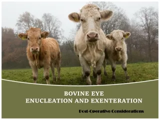
Introduction into the Bovine eye and diagnostic and surgical procedures
Vision is essential for food- or fiber-producing animals to safely exist in their environments and compete for food. Ocular diseases can result in considerable discomfort to the patient with resultant poor weight gain, decreased milk production, behavioral problems, and poor performance.
A complete history and physical examination should be performed on all patients, even if the complaint concerns “just” the eye. • Many ocular problems in food animal species are manifestations of systemic diseases; and these disorders should be ruled out before the ocular examination commences because dehydration, anaemia, icterus, and others of these types of physical parameters may affect the ocular findings.
Historical information should include the owner’s or herdsman’s assessment of the patient’s visual status. • Careful observation of the patient may be necessary if normal vision is questioned and should be performed on a herd animal patient in separate and unfamiliar surroundings. • Head carriage should be noted (visually deficient animals often carry their heads close to the ground).
Previous ocular diseases should be queried. Current and previous ocular and systemic medication including conventional and alternative therapeutic modalities should be noted. • Whenever possible, examination of the eyes should be performed in a quiet, dark room. • Adequate restraint of the head is essential, and sedation of the patient may be necessary. In addition to an ophthalmoscope, minimal specialized equipment is necessary.
Intraorbital structures • Globe, nerves (II-VII), blood vessels, gland of the 3rd eyelid, lacrimal gland, zygomatic salivary gland, extraocular muscles, masticatory muscles, fat, and periorbita. • Bone and sinuses incompletely surround the orbital soft tissues in domestic animals. • Alterations in any of the bony or soft tissues also alters the remaining tissues and usually the position of the eye.
Clinically significant Formina and Fissures of the Orbit • Optic foramen - optic nerve (II). The dura of the optic nerve is continuous with the brain's dura so injections into the nerve can affect the brain. • Foramen orbitorotundum (cow, sheep, pig) - III, IV, V (both parts), VI, and maxillary artery. This is the site of the Peterson nerve block in cattle for enucleation.
Eyelids • Cattle have three eyelids. • The upper and lower lids help to protect the eye from the environment, distribute tears over the entire eye surface, and control the amount of light that enters the eye. • The third eyelid is located in the inner corner of the eye and sweeps across the eye as it closes. • It functions to protect and lubricate the eye. • It has its own set of tear glands that produce lubricating tears for the entire eye.
Problems with the lids can result in pain, swelling, redness, excessive tearing, and drainage from the eye. • Animals with lid pain may attempt to rub their eyes, squint, and show other signs of pain. • Problems with the lids can lead to additional problems with closely associated structures such as the cornea, conjunctiva, and nasolacrimal drainage system of the eye.
DIAGNOSTIC PROCEDURES Always strive for an accurate diagnosis prior to Rx.
Physical Examination - look, feel, retropulse the globe, open the mouth. • Pain on opening the mouth is typical of inflammatory orbital disease, whereas neoplasia tends to be nonpainful. • This is the cheapest, most effective diagnostic procedure and must be performed thoroughly in order for further diagnostic tests to be appropriately selected. https://www.youtube.com/watch?v=A3r3MI_HBIw
Radiography – Take films before invading the orbit to avoid confusing artefacts. Plain films are useful if bony or sinus disease is suspected. Can be difficult to interpret. - Usually need general anesthesia. Plain films rarely give an exact diagnosis but can be very helpful in suggesting a prognosis. (Bony involvement suggests a poorer prognosis). • B scan Ultrasonography • Do before invading the orbit. Helpful in soft tissue disease and in guiding a fine-needle aspirate of a small or localized lesion. Does not image bone well. • Does not require general anesthesia. With experience the soft tissue images can be mentally combined with radiographs to better characterize the orbital disease.
Aspiration and cytology • An aspirate or biopsy is usually required to get an accurate diagnosis in orbital disease. • Disadvantages are possible damage to important orbital structures (optic nerve), infection, failure to indicate the full extent of the pathology, the need for anesthesia or sedation (some patients), and getting an unrepresentative sample. • Usually failure to yield a diagnosis is because the lesion is small or localized, unrepresentative tissue was sampled, or lack of tissue architecture to aid in diagnosis. • Neoplasia can be missed if only the necrotic center of a tumor is aspirated.
Surgical Exploration of the Orbit - Necessary if less invasive techniques fail to yield a diagnosis. It allows you to fully assess the extent of the disease, it can be therapeutic as well as diagnostic, and provides representative tissue for pathology. -Disadvantages include general anesthesia, the need for advanced surgical training, infection, potential disfigurement or damage to vital structures, time involved and the expense.
Ophthalmic surgery • In large animals is nearly always related to the economic value of the animal, the amount of post operative treatment and the type of lesion. • Especially in cattle the majority of ophthalmic surgery is of the peri-ocular type and includes procedures involving the orbit, eyelids, nictitating membrane , nasolacrimal system and conjunctiva.
Types of orbital surgery • Enucleation With or Without an Orbital Prosthesis - Removal of only the globe. A silicone intraorbital prosthesis can be used to improve cosmesis. A prosthesis is contraindicated in the presence of orbital infection or neoplasia. https://www.youtube.com/watch?v=UPorygigmjo • There are two types : • Subconjunctival – Used in non-contaminated cases (infectious/ neoplasia confined to the globe), the eyelid remains open • Transpalpebral – Removes the globe, short piece of the optic nerve, lid margins, conjunctiva, third eyelid and gland of third eyelid
Indications for enucleation • Squamous cell carcinoma • Lymphosarcoma • Chronic glaucoma • Prolapsed retrobulbar fat (Phthisis bulbi) • Severe trauma or proptosis. • Rupture • Chronic endopthalmitis or panopthalmitis • Retrobulbar abscessation; periorbital cellulitis • Perforating ulcers • Lacerations, orbital fractures, foreign bodies
Exenteration With or Without an Orbital Prosthesis - The removal of all of the orbital contents and the globe. • Usually performed for orbital neoplasia or infection. An orbital prosthesis can be implanted if there is no residual neoplasia or infection. https://www.youtube.com/watch?v=n9Fv-vDC2HI SCC - Eye enucleation in cow
Evisceration and Intraocular Prosthesis - Removal of the intraocular contents leaving the corneo-scleral shell that is then filled with a silicone prosthesis. Contraindicated in the presence of intraocular infection, concurrent ocular disease such as corneal ulceration or dry eye, and ocular neoplasia. More cosmetic than an enucleation. https://www.youtube.com/watch?v=tu6IU3Ra3eM • Extrascleral Prosthesis (shell) - This is the typical artificial eye used in humans and is rarely performed in veterinary ophthalmology due to difficulties in maintaining the prosthesis in place and its expense. Occasionally done in show horses.
Ophthalmic surgery in large animals is performed under topical, regional or general anesthesia. • In cattle, regional and general anesthesia are the most frequently used. Both methods need sedation prior to induction of anesthesia. • General anesthesia can be obtained by using chloral hydrate or barbiturates. • For regional anesthesia we can use the combined retrobulbar and auriculopalpebral nerve block.
Today’s laboratory procedures • Pre- surgical procedure of the eye • Administration of nerve blocks: • Auriculopalpebral • Petersen’s • 4 point Retrobulbar • Subconjunctival injection ( used when treating Pink eye) • Removal of the third eyelid • Exenteration and Transpalpebral enucleation of a bovine cadaver’s eye • Surgical procedure for closure of the orbital region • Discuss post surgical care and complications