Download
1 / 25
270 likes | 579 Vues
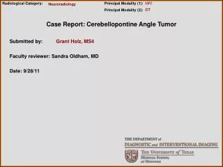
Radiological Category:. Principal Modality (1): Principal Modality (2):. MRI. Neuroradiology. CT. Case Report: Cerebellopontine Angle Tumor. Submitted by:. Grant Holz, MS4. Faculty reviewer: Sandra Oldham, MD. Date: 9/28/11. Case History.

E N D
Radiological Category: Principal Modality (1): Principal Modality (2): MRI Neuroradiology CT Case Report: Cerebellopontine Angle Tumor Submitted by: Grant Holz, MS4 Faculty reviewer: Sandra Oldham, MD Date: 9/28/11
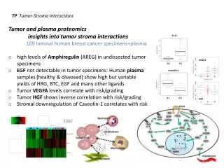
Case History 65 yo female with PMH of a left-sided cerebellopontine angle tumor resected in 1999 presented with a 2 week history of headache, dizziness, and N/V. She went to an outside hospital where a CT scan showed a new left-sided cerebellopontine angle mass, and she was transferred to Memorial Hermann Hospital. She was admitted to the neurosurgery ICU and IV steroids were started. An MRI was obtained which showed a large enhancing heterogeneous mass, measuring 4.6 x 4.4 x 4.0 cm situated in the left cerebellopontine angle. The patient remained at GCS 15 with no clinical signs of hydrocephalus. Her neuro exam demonstrated left-sided weakness and left facial palsy that were secondary to her left posterior craniotomy in 1999, and have been stable since. Due to the large complexity of the tumor, it was decided to conservatively manage the patient on an outpatient basis, in order to explore surgical options.
T1 Axial Post-Contrast Slice # 1 (Superior margin of tumor)
T1 Axial Post-Contrast Slice #2
T1 Axial Post-Contrast Slice #3
T1 Axial Post-Contrast Slice #4
T1 Axial Post-Contrast Slice #5
T1 Axial Post-Contrast Slice #6
T1 Axial Post-Contrast Slice #7 (Inferior margin of tumor)
Background • Cerebellopontine angle anatomy • - Located in posterior fossa • Bounded by: • brainstem medially • petrous portion of temporal bone laterally • middle cerebellar peduncle superiorly • arachnoid tissue of lower CN inferiorly Symptoms of Cerebellopontine angle masses - Sensory hearing loss - Disequilibrium or vertigo - Hemifacial weakness or paralysis - Decrease in sensation of the external auditory canal - Wide based gait - Headaches and visual loss Study of choice = MRI w contrast
Test Your Diagnosis Which one of the following is your choice for the appropriate diagnosis? • Vestibular schwannoma / Acoustic neuroma • Meningioma • Epidermoid tumor
Enhancing heterogeneous mass in the L CPA measuring 4.6 x 4.4 x 4 cm w multiple areas of cystic degeneration and hemorrhage. An intracanalicular component is noted with widening of the L internal auditory canal. Marked mass effect upon the adjacent brainstem and cerebellum w stretching and thinning of the L middle cerebellar peduncle and edema in the L cerebellar hemisphere. The 4th ventricle is effaced and there is mild supratentorial hydrocephalus with transependymal CSF flow. The mass extends into the L jugular foramen. The L jugular vein is completely effaced in the foramen and is decreased in caliber just below the skull base with filling via collaterals and the inferior petrosal sinus. The mass also extends inferiorly to the foramen magnum on the left displacing the cervical medullary junction towards the right. The L sigmoid sinus is decreased in caliber, but enhances on the Stealth suggesting at least partial patency. Findings: Differentials (for lesions arising within the CPA): • Vestibular schwannoma / Acoustic neuroma • Meningioma • Epidermoid tumor • Less likely: Arachnoid cyst, Non-acoustic cranial nerve schwannomas, Vascular malformations, Dermoids, Teratomas, and Lipomas.
Acoustic neuromas Most common CPA lesion Middle to late decades of life Usually unilateral Benign and slow growing Characteristic features include: centered at porus acousticus acute angle to petrous bone involvement of IAC no dural tail no calcifications lack prominent vasculature never penetrate into middle cranial fossa Homogenous mass T1: isointense to brain and hyperintense to CSF T2: hyperintense to brain and iso/hypointense to CSF on T2 Intense enhancement on T1 post contrast Discussion
Meningiomas 2nd most common CPA lesion Characteristic features: Obtuse angles to petrous bone Do not extend into IAC Often have dural tail Commonly demonstrate calcification May show flow voids of pial blood vessels Heterogeneous masses w possible central clearing T1: isointense to brain T2: between brain and CSF in intensity Post contrast: not as intense enhancement as acoustic neuroma Discussion
Epidermoid tumors 3rd most common CPA lesion Congenital lesions Present in adulthood Stratified squamous epithelium which surrounds a mass of keratin debris Benign and slow growing Facial weakness, paralysis, and spasm Expand into nearby structures Homogenous lesion T1: very low intensity, isointense to CSF T2: very bright, isointense to CSF Discussion
AcousticNeuroma Diagnosis
1. Bailey, Byron J. Head and Neck Surgery – Otolaryngology. Lippincott. New York, NY. 2001.2. Brackmann, Shelton, Arriaga. Otologic Surgery. W.B. Saunders Company, New York. 2001.3. Fisch, Mattox. Microsurgery of the Skull Base. Georg Thieme. New York, NY. 1988.4. Lang, Johannes. Clinical Anatomy of the Posterior Cranial Fossa and its Foramina. Thieme Medical Publishers, Inc. 1991.5. McElveen, Dorfman. “Petroclival Tumors” Otolaryngology Clinics of North America. 2001, 34: 1219-1230.6. Mendenhall, et al. “Management of Acoustic Neuroma” American Journal of Otolaryngology. 2004; 25: 38-47.7. Myers, et. al. Operative Otolaryngology. Head and Neck Surgery. Saunders Company. Philadelphia, PA. 1997.8. Som, Curtin. Head and Neck Imaging. Mosby. St. Louis, MO. 2003. References