Download
1 / 41
410 likes | 446 Vues
Explore the definition and overview of angiogenesis, endothelial cell migration, and tube formation. Learn about the sprouting process, VEGF pathway, associated diseases, tumor growth, and angiogenesis assays. Discover activators, inhibitors, and the role of VEGF in blood vessel growth.

E N D
P02_Endothelial cells: Functional Analysis - Migration-Tube formation ANGIOGENESIS Endothelial Cell Migration Assay Endothelial Cell Tube Formation Assay
P02_Endothelial cells: Functional Analysis - Migration-Tube formation CONTENT • Angiogenesis: Definition & Overview • Angiogenesis: Assays Overview • Endothelial Cell Migration Assay (using FluoroBlokTM Insert system) • Endothelial Cell Tube Formation Assay
P02_Endothelial cells: Functional Analysis - Migration-Tube formation Angiogenesis: Definition • VASCULOGENESIS • embryonic formation and differentiation of the vascular system • formation of new blood vessels from angioblasts or progenitor stem cells (de-novo vessel synthesis) • ANGIOGENESIS • formation of new blood vessels (sprouting, branching etc.) from existing vessels
P02_Endothelial cells: Functional Analysis - Migration-Tube formation Angiogenesis: The Sprouting Process • TRIGGERS FOR SPROUTING • Infections • Tissue damage • Tumors • Hypoxia • SPROUTING SIGNALS • Mainly VEGF and HIF • SPROUTING EVENTS • Secretion of proteases (MMPs) • Digestion of basal lamina of vessels • Migration to signal source • Endothelial cell proliferation • Tube formation new vessels bring O2 and reduce sprouting trigger
Endothelial cells as target for drug screening DRUG e.g. Tyrosin kinase inhibitors TKI “mode of action” Inhibition of prolfieratio- Migration-Vessel formation? AR? 5
P02_Endothelial cells: Functional Analysis - Migration-Tube formation Angiogenesis: Activators and Inhibitors
P02_Endothelial cells: Functional Analysis - Migration-Tube formation Angiogenesis: The VEGF Pathway
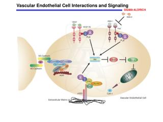
P02_Endothelial cells: Functional Analysis - Migration-Tube formation Angiogenesis: VEGF as a Key Factor • Vascular Endothelial Cell Growth Factor (VEGF) • Secreted homodimeric growth factor • Expressed by a variety of vascularized tissue • Different isoforms. VEGF165 as key regulator of blood vessel growth • VEGF receptors • VEGF-R1, VEGF-R2, VEGF-R3 • VEGF-R2 as major mediator of EC proliferation, migration, angiogenesis • VEGF released from tumors • Potently induces angiogenesis in vivo • Induced vasodilation / vascular permeability / endothelial cell (EC) fenestration • ECs in new tumor vasculature display VEGF dependence • Promotes EC survival, proliferation and migration • Clinical angiogenic inhibitors • Bevacizumab (Avastin) (antibody against VEGF) • PTK787, ZD6474, SU6668, SU11248 (inhibits VEGF-R2)
P02_Endothelial cells: Functional Analysis - Migration-Tube formation Angiogenesis: Associated Diseases
P02_Endothelial cells: Functional Analysis - Migration-Tube formation Angiogenesis: Tumor Growth
P02_Endothelial cells: Functional Analysis - Migration-Tube formation Angiogenesis is sexy…
P02_Endothelial cells: Functional Analysis - Migration-Tube formation ANGIOGENESIS ASSAYS OVERVIEW
P02_Endothelial cells: Functional Analysis - Migration-Tube formation Organ Culture: Rat Aortic Ring Assay • slices of vessels are incubated on Matrigel • sprouting of vessels starting from the • endothelial layer of vessel
P02_Endothelial cells: Functional Analysis - Migration-Tube formation In Vivo Assays: CAM & Matrigel Plug Chick chorioallantoic membrane (CAM) assay: Angiogenic factors are administered Onto the allantoic membrane of the chicken Embryo and angiogenesis is analyzed Matrigel Plug Assay: A chilled mixture of cells/Matrigel HC is injected Subcutaneously. In the body, the plug will be broken down over Time (10-14 d) Invasion of vessels into plug starts at day 2-3
P02_Endothelial cells: Functional Analysis - Migration-Tube formation Angiogenesis: Endothelial Cells Commercially available endothelial cells come from many sources: HUVEC: Human Umbilical Vein Endothelial Cells* HMEC: Human dermal MicrovascularEndothelial Cells Capillary EC, easily accessible human material BAOEC: BovineAOrticEndothelialCells Bovine sources are cheap Different cell lines may respond differently in assays max. life cycle of EC is around 10-20 passages; thereafter, cells lose phenotype and die We use passage 4
P02_Endothelial cells: Functional Analysis - Migration-Tube formation HUVEC: Material – Isolation-Characterization-long term stability
P02_Endothelial cells: Functional Analysis - Migration-Tube formation • Procedure: • Consent of the mother (+ Ethic commission) • Length of the cord 20-30 cm • Store immediately after birth in medium (RPMI 10% FCS + AB) • Subsequent Rrinsing with Heparin to avoid blood coagulation not possible • Transfer to the lab as outlined in PPP 01 • Storage at 4°C over night • Rinsing the vein using RPMI Medium • If blood is washed out: • Digest the inner endothelial cell monolayer by using one of these techniques • Control the absence of smooth muscle cells and fibroblast (prolonged digestion)
Isolation and propagation in specific medium • Microvascular EC • Tumor tissue • Foreskin • Using enzymatic digestion and selection by any technique • Bovine/swine aorta • Abrasion of the EC Monolayer Monolayer Cell Isolation - 2002
EC-quality control • Material: • Human EC derived from UC from healthy individuals • Enzymatic digestions as outlined • Cultivation in EGM-2 (Lonza) upt to 12th passage • Control by differentiation (vw-factor) and function • Freezing using PC-control device • Long term storage in Nitrogen • Thawing and quality control (WST, BrdU, Immunostaining vWF) • Upon release use in sreening/research HU 73 p6 Phasenkontrast HU 74 p5 vWF Immunfärbung HU 106 p7 Phasenkontrast 19
Quality control during serial passages • Cultivation of different batches up to passage 10 • Documentation of differentiation by Immunostaining • Quantifiation of vWF in % of total cells 20
P02_Endothelial cells: Functional Analysis - Migration-Tube formation Functional Analysis • Differentiation • Growth behavior • Migration • Tube (vessel) formation • Behavior under flow • 3D Models
HUVEC 73 Migrationsassay • Cells seede on 12 well MTP • Scratch after reaching confluence 4d • Microscopic control of EC migration into the “wound” Wound= Scratch control 2 h control 24 + 10µM /L Drug 24h 22
P02_Endothelial cells: Functional Analysis - Migration-Tube formation Cell migration (Scratch Assay) • Only horizontal • No Gradient of growth factors • Quantification by cell counting/imaging • Ready-to-use plates available ( expensive) Alternative: Boyden chamber assay – vertical from top to bottom
P02_Endothelial cells: Functional Analysis - Migration-Tube formation ENDOTHELIAL CELL MIGRATION ASSAY
P02_Endothelial cells: Functional Analysis - Migration-Tube formation In vitro Assays: EC migration and invasion
P02_Endothelial cells: Functional Analysis - Migration-Tube formation Migration / Invasion Assays: the hard & the smart way…
P02_Endothelial cells: Functional Analysis - Migration-Tube formation Principle Of Cell Migration Assay
P02_Endothelial cells: Functional Analysis - Migration-Tube formation Compatible Fluorophores for BD FluoroBlokTM Insert Systems
P02_Endothelial cells: Functional Analysis - Migration-Tube formation Endpoint or Real time kinetic Assay
P02_Endothelial cells: Functional Analysis - Migration-Tube formation Real-Time Analysis of HUVEC Migration
P02_Endothelial cells: Functional Analysis - Migration-Tube formation Correct coating may be critical for cell migration
P02_Endothelial cells: Functional Analysis - Migration-Tube formation Timecourse of Chemotaxis in Monocytic Cells • Response of Macrophage Chemoattractant Protein 1 (MCP-1)-induced chemotaxis in monocytic cells: • Calcein-prelabeled cells (top chamber) were incubated with 25 nM MCP-1 (bottom chamber) • Bottom fluorescence was measured at varying time points • Data on the graph are representative for a typical experiment • 20-25 min incubation is optimal
P02_Endothelial cells: Functional Analysis - Migration-Tube formation Importance of Optimizing Cell Seeding Density
P02_Endothelial cells: Functional Analysis - Migration-Tube formation ENDOTHELIAL CELL TUBE FORMATION ASSAY
P02_Endothelial cells: Functional Analysis - Migration-Tube formation EC Tube Formation: Assay Features • PRINCIPLE • ECs build up tube networks when cultured under appropriate conditions • labeled tube networks will be Calcein-stained and • tube length be measured using automated fluorescence microscopy • RAPID DATA • tube formation & assay data after 24 hours from seeding
P02_Endothelial cells: Functional Analysis - Migration-Tube formation EC tube formation assay EC are cultivated at desired cell density onto the top of high concentrated Matrigel (4-8 mg/ml Any drug/growth factors can be applied into the gel Monitoring of “branches” by eye or imaging software
P02_Endothelial cells: Functional Analysis - Migration-Tube formation EC Tube Formation: 96 well image acquisition
P02_Endothelial cells: Functional Analysis - Migration-Tube formation EC Tube Formation: Number of seeded cells is critical • cell lines differ in the number of cells that is optimal for tube formation • seeding cell density will result in cell growth as a layer rather than as tubes • The optimal cell seeding number should be titrated cell-line specifically
P02_Endothelial cells: Functional Analysis - Migration-Tube formation EC Tube Formation: Data consistency of inter-day experiments
P02_Endothelial cells: Functional Analysis - Migration-Tube formation EC Tube Formation: Influence of Compounds II
IBIDI-Kammer 1 4 5 2 6 3 FLOW-orientiation 1 – 6 = imageposition P02_Endothelial cells: Functional Analysis - Migration-Tube formation Subcultivation into Flow-chamber: BdSMC, p11, 07.04.09Flow-Start: 08.04.09FLOW: 0,30 ml/minShear-Stress: 1,54 dyne/cm2Coating:uncoated Bild: 10.04.09 Control w/o flow (Start) 3 4 Smooth muscle cells under shear stress 5 6