Cell Migration
Cell Migration. IBS-501, April 16, 2001 Barry D. Shur. Epithelial-mesenchymal transformation. Basal lamina controls epithelial phenotype Degradation of basal lamina leads to loss of cell polarity and mesenchyme transformation Collagen gels vs basal lamina

Cell Migration
E N D
Presentation Transcript
Cell Migration IBS-501, April 16, 2001 Barry D. Shur
Epithelial-mesenchymal transformation • Basal lamina controls epithelial phenotype • Degradation of basal lamina leads to loss of cell polarity and mesenchyme transformation • Collagen gels vs basal lamina • Anti-E-cadherin antibodies break adhesions and lead to mesenchyme transformation
What induces transformation? • TGFß induces loss of E-cadherin, increase in fibronectin synthesis, transformation • Scatter factor/hepatocyte growth factor, binds to c-met (tyrosine kinase) and induces epithelial dissociation • Metalloprotease degradation of basal lamina
Biology of Cell Migration • Polarized insertion new membrane (with receptors) • Generate an intracellular force • Dissociation from substratum
Polarized insertion of new membrane • In response to stimuli, MTOC reorients along with Golgi • Enables membrane insertion into leading edge
Polarized insertion of membrane glycoproteins • Vesicular stomatitis virus G protein in ER at non permissive temp, shift to permissive temp, G protein moves through Golgi and inserted into leading lamellipodia • Microtubule disrupting drugs (nocodazole) inhibit Golgi reorientation, membrane (G protein) inserted uniformly or randomly
Generating an intracellular force • Lamellipodia projections need tension, infrastructure • Actin filament polymerization generates force • Cytochalasin B caps free end of F-filaments, inhibits lamellipodia formation/migration • Microinjected actin incorporated into leading edge, snd moves rearward • Photobleach spot of rhodamine-conjugated actin moves rearward
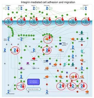
Polarized insertion of membrane glycoproteins • Integrins inserted into leading edge • binds ligands (I.e., fibronectin) • ligand-induced clustering, • activates FAK, • recruits linker proteins to polymerize actin
Integrin clustering activates FAK and recruitment of cytoskeleton components
Actin polymerization at the leading edge
Actin filaments terminate in focal adhesions
How do cells release from the substratum?