Download
1 / 12
130 likes | 342 Vues
Cell migration: Rho GTPases lead the way. Xia Fan. How migration occurs?.

E N D
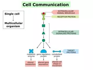
How migration occurs? • The major driving force of migration is the extension of a leading edge protrusion or lamellipodium, the establishment of new adhesion sites at the front, cell body contraction, and detachment of adhesions at the cell rear.
The Rho GTPase switch(Rho,Rac,Cdc42) • Cycle between a GDP-bound, inactive form GTP-bound, active form. • The cycle is regulated by • GEF:promote the exchange of GDP for GTP • GAP:enchance the intrinsic GTPase activity • GDI:block the cycle by sequestering and solubilizing he GDP-bound form • Predominantly through GEF. • How they regulate migration: • Rho:assembly of contractile actin:myosin filaments • Rac:polymerization of actin to form lamellipodial • Cdc42:polymerization of actin to form filopodial protrusion • all promotes the assembly of integrin-based, matrix adhesion complex
Regulation of Rho GTPases during cell migration • Observation: Rac can be visualized with highest concentrations at the leading edge. • Focus: Rac activation-two pathways
PI-3-kinase and PI(3,4,5)P3 • Extracellular signal activated receptor protein activated PI-3-kinase PI(3,4,5)P3 docked Rac GEF • How Rac GEF promotes GTP loading • PI(3,4,5)P3 binding relieves interaction between PH and DH domain to stimulate activity • Loss of activity due to deletion of PH domain is restored by addition of a CAAX motif to stable the binding.(The DH domain induces Rac to displace GDP and bind to GTP. The DH domain is invariably proceeded by a PH domain, which greatly increase catalytic efficiency.) • PI(3,4,5)P3 regulates PIX, which activates Rac and interacts with PAK • Rac activation stimulates PI 3-kinase, resulting a positive feedback loop
p130Cas/CrkII/DOCK80 • Extracellular signal activated receptor protein activated PI-3-kinase PI(3,4,5)P3 docked DOCK180 • How DOCK180 promotes GTP loading • Bind to the adaptor protein Crk, which in turn associates with another adapor p130Cas. The Crk/p130Cas/DOCK180 can lead to Rac activation. • Two plausible explanation of the biochemical mechanism: • DOCK180 recruits a DH-containing GEF to the complex. • The C-terminus of DOCK180 can stimulate GTP loading on Rac. • The basic region of DOCK180 seems to gunction in the same way as the PH domain of DH-domain-containing GEF
Downstream effects of Rho GTPases during cell migration • Regulation of the actin cytoskeleton • Rac: generates a protrusive force through the localized polymerization of actin. • Cdc42: generates filopodia through the localized polymerization of actin. • Rho: generates focal adhesion and cell contractility by across-linking actin:myosin filaments. • Regulation of the microtubule and polarity • Efficient and persistent long-range migration requires stabilizeion of cell polarity and is achieved through reorganization of the microtubule cytoskeleton. • Cdc42 plays a crucial role in polarized migration by reorientation of microtubules and centrosome.
Regulation of the actin cytoskeleton • Either Rac or Cdc42 can activate p65PAK; Cdc42 activates WASp and N-WASp;Rac activates the Scar/WAVE. • p65PAK 1.regulates focal adhesion turnover with help of PIX and GIT1(mechanism is unkonwn) 2.phosphorlates and activates LIMK, which in turn phosphorylates and inactivates cofilin.(confilin faciliates subunit dissociation from the pointed end of actin filaments and induces filament severing and is essential fro promoting filament treadmilling at the front) • WASP/SCAR/WAVE 1.stimulates the Arp2/3 complex, which can initiate actin polymerization. 2.WASP/WAVE can bind to profilin, which acts synergistically with Arp2/3 to speed up actin polymerization.
Regulation of the actin cytoskeleton • Rho can activates p160ROCK and mDia • p160ROCK 1.works like p65PAK,leading to stabilization of actin filaments within actin:myosin filament bundles. 2.interacts with and phosphorylates the myosin binding subunit of myosin light chain phosphatase and inactivates it,leading to increased levels of myosin phosphorylation, which then can cross-link actin filaments. • mDia is linked to actin filament assembly. (mechanism is unknown)
The microtubule cytoskeleton and polarity • Rho:activates mDia, which directly interacts with microtubules and promotes their capping, leading the stabilization of microtubules. • Rac:promotes microtubule elongation through 1. p65PAK-dependent phosphorylation by activating p65PAK 2.inactivating stathmin, the microtubule destabilizing protein. • Cdc42 can activates the complex Par6/PKC. PKCphosphorylates and inactivates GSK-3. This induces the association of APC with the plus ends of microtubules specifically at the leading edge. Through a dynein-or dynactin-dependent mechanism, this results in microtubule reorganization and centrosome reorientation. (APC could also serve to localize Asef, a Rac-specific GEF, to sites of Rac-dependent actin polymerization)