Biomimetic Conducting Polymers
Craig Milroy ( ChE ). Dr. Zin Khaing Post Doc (Neurobiology). E-mail: schmidt@che.utexas.edu Office: BME 4.202l Lab: BME 4.318 Phone: 512- 471 -1690. ELECTROACTIVE MATERIALS. NATURAL MATERIALS. Biomimetic Conducting Polymers

Biomimetic Conducting Polymers
E N D
Presentation Transcript
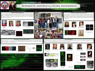
Craig Milroy (ChE) Dr. ZinKhaing Post Doc (Neurobiology) E-mail: schmidt@che.utexas.edu Office: BME 4.202l Lab: BME 4.318 Phone: 512- 471 -1690 ELECTROACTIVE MATERIALS NATURAL MATERIALS Biomimetic Conducting Polymers Our lab is developing advanced electronic materials specifically tailored and optimized for biomedical applications. The biomedical applications currently receiving the most attention are drug delivery devices, probes to record/stimulate the activity of brain cells, and tissue scaffolds for tissue engineering(i.e. the replacement or restoration of damaged or malfunctioning biological tissues) in the exciting field of artificial/prosthetic limbs/organs. Regeneration in the Central nervous system Our research interests are in the field of plasticity and regeneration within the central nervous system. Currently, we are using rodent spinal cord injury as a model system to study how engineered materials aid in the process of axonal regeneration. Projects include acellular nerve grafts, hyaluronic acid-based scaffolds for axonal regeneration within the adult mammalian spinal cord, and 3D in vitro culture systems using hyaluronic acid-based hydrogels for neural progenitor cell differentiation. Dr. John Hardy Post Doc (Chemistry) Chase Cornelison (ChE) Phase contrast (left) and fluorescent (right) microscopy images of schwann cells cultured at a conductive/nonconductive (top/bottom) substrate interface Myelin-Derived inhibitors Buffer SEM of polypyrrole-coated PLGA fibers. Neurons extend longer, more oriented axons with combined physical and chemical cues Attenuating inflammatory wound-healing response through bilayer, bifunctional HA materials Eric Spivey (BME) Co-advisor: J. Shear Sarah Mayes (BME) Derek Hernandez (ChE) Co-advisor: J. Shear Richelle Thomas (ChE) Myelin-Derived inhibitors + Soluble NgR Myelin-Derived inhibitors + Clone 40 SEM micrograph of a Schwann cell cultured on polypyrrole doped with polystyrene sulfonate. Aptamers directed against NgR can limit myelin-derived inhibition of neurite outgrowth in cultured DRG neurons Hieu Nguyen (BME) After cervical spinal cord injury, rats show functional recovery in the cylinder Ryan Nagao (BME) Collaborator: L. Suggs Biotinylateddextran amine anterograde tracing from the motor cortex through the cortical spinal tract of the spinal cord after injury Schwann cells grown on nerve growth factor coated wells with electrical stimulation Sydney Geissler (BME) Mechanisms of Cell-Materials Interactions The goal for this aspect of our research is to use experimental approaches, theoretical modeling and computational simulation (in collaboration with others) to better understand how cells and biomolecules interface with materials (e.g. affinity peptide binding and Schwann cell migration on conducting polymers). Natural Biopolymers Our group is utilizing novel techniques to process biopolymer based materials (e.g. hyaluronic acid) into forms to be used in therapeutic applications. In a parallel approach, we are investigating decellularized tissues for use as regenerative scaffolds. Dr. Scott Zawko Research Associate (ChE) Glial cells do not adhere to pyrrole-HA coated electrodes Cells present specific responses to different substrates A) SELECTED PUBLICATIONS Lee, J.Y., C.A. Bashur, C.A. Milroy, L. Forciniti, A.S. Goldstein, C.E. Schmidt (in press). Nerve growth factor-immobilized electrically conducting fibrous scaffolds for potential use in neural engineering applications. IEEE Transactions on NanoBioscience. Suri, S., L.-H.Han, W. Zhang, A. Singh, S. Chen, C.E. Schmidt (2011). Solid Freeform Fabrication of Designer Scaffolds of Hyaluronic Acid for Nerve Tissue Engineering. Biomedical Microdevices. 13(6):983-93. Broda, C.R., J.Y. Lee, S. Sirivisoot, C.E. Schmidt, B.S. Harrison (2011). A Chemically Polymerized Electrically Conducting Composite of PolypyrroleNanoparticles and Polyurethane for Tissue Engineering. Journal of Biomedical Materials Research Part A. 98:509-16. Nagao, R.J., S. Lundy, Z.Z Khaing, C.E Schmidt (in press). Functional Characterization of Optimized Acellular Peripheral Nerve Graft in a Rat Sciatic Nerve Injury Model. Neurological Research. Forciniti, L., N.K., Guimard, S. Lee, C.E. Schmidt (2010). Unique electrochemically synthesized polypyrrole:poly(lactic-co-glycolic acid) blends for biomedical applications. Journal of Materials Chemistry. 20: 8865–8874. [Journal Impact Factor= 4.795; 2009] Seidlits, S.K., Z.Z. Khaing, R.R. Petersen, J.D. Nickels, J.D. Vanscoy, J.B. Shear, C.E. Schmidt (2010). The effect of hyaluronic acid hydrogels with tunable mechanical properties on the differentiation of neural progenitor cells. Biomaterials. 31: 3930 - 3940 Zawko SA, Schmidt CE (2010). Simple benchtop patterning of hydrogel grids for living cell microarrays. Lab Chip. Feb 7;10(3):379-383. Seidlits, S.K., C.E. Schmidt, J.B. Shear (2009). High-Resolution Patterning of Hydrogels in Three Dimensions using Direct-Write Photofabrication for Cell Guidance. Advanced Functional Materials. 19:3543-3551. Cells migrate down an IKVAV-functionalized 3D structure in a hyaluronic acid hydrogel B) Electron micrographs of vascular casts A cell migrating toward an immobilized structure Photoacoustic image (750 nm) of decellularized construct after addition of nanotracer-containing macrophages C) Chemical gradients immobilized on protein microarchitectures 25 µm GMHA hydrogelstemplated with A) potassium dihydrogen B) guanidine C) urea 10 µm Hydrogels patterned with a fibrillar microstructure