Mitosis: A Comprehensive Lesson on Cell Division
410 likes | 638 Vues
Master the significance of mitosis in life cycles, cell cycle regulation, and chromosomes through detailed explanations and interactive activities. Explore the stages of mitosis, genetic duplication, and cellular checkpoints with engaging animations and practical demonstrations.

Mitosis: A Comprehensive Lesson on Cell Division
E N D
Presentation Transcript
Learning Objectives You will have been successful if by the end of the lesson you can demonstrate and apply your knowledge and understanding of • The significance of mitosis in life cycles To include growth, tissue repair and asexual reproduction in plants, animals and fungi. • Cell cycle To include the processes taking place during interphase (G1, S and G2), mitosis and cytokinesis, leading to genetically identical cells. • How the cell cycle is regulated To include an outline of the use of checkpoints to control the cycle
The beginning……. Ovum Its nucleus contains 23 chromosomes Zygote The nucleus from the sperm fuses with the ovum nucleus – now there are 46 chromosomes in the cell (23 pairs) Millions of cells will be produced from this beginning + Sperm Its nucleus contains 23 chromosomes
Karyotypes The chromosomes from a dividing cell can be photographed and organised into the pairs – one from the mother and one from the father Each pair has a characteristic size, shape and banding pattern (except for the sex chromosomes where the X and Y chromosomes are different) Karyotype from a female Autosomes 22 pairs Make a karyotype Try it yourself Heterosomes 1 pair
Homologous chromosomes • The 22 pairs of autosomes are known as homologous chromosomes – they have the same genes in the same places
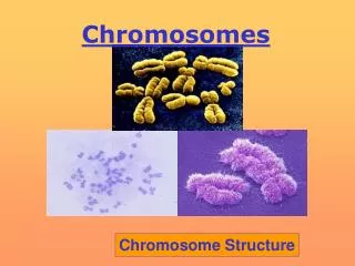
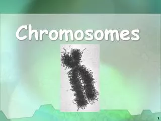
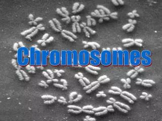
Visible chromosomes? • When we can see distinct chromosomes its because the DNA molecules are tightly coiled About 3% of one of the DNA molecules of one of the 46 chromosomes ! Packing this amount into the nucleus takes some doing……
The solution….. • Coil the DNA strand around histone proteins • Keep on coiling…… DNA coiling animation
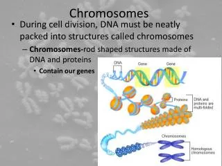
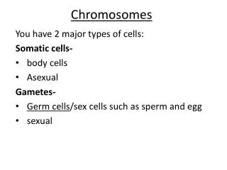
Prokaryotes use a form of cell division called binary fission because they don’t have a nucleus and therefore cannot carry out mitosis Significance of Mitosis Mitosis is the division of the nucleus • Daughter cells are identical to the parent cell • Mitosis occurs during • Growth • Repair of tissues • Asexual reproduction
Why the X-shape ? • Use your materials to create a “cell” with 2 homologous pairs, 4 mitochondria (pasta) and 6 ribosomes (beans). Now create two identical cells. Paternal chromosomes Maternal chromosomes Homologous pair 1 Homologous pair 2 How does the cell produce the extra DNA? What else would the cell have to do…..? Can you explain this diagram?
Chromatids • Each molecule of DNA in the nucleus replicates to produce 2 identical strands • The strands are called chromatids • They remain joined together at the centromere
So…before the cell can begin to divide • DNA molecules must replicate • And then coil and coil again and again… • The number of organelles needs to increase
McGraw Hill Animation Cell Cycle Interphase G1 – protein synthesis and growth S – replication of DNA G2 – organelles divide, DNA condenses Mitosis Nuclear division – 4 stages Cytokinesis Animation re p53 and DNA proof reading And lung cancer Cell divides into two Nobel Prize game re cell cycle and checkpoints
Cell cycle and Mitosis (Cells Alive) Cell Cycle Checkpoints These control the cell cycle. Ensure that each stage is completed properly before entering the next stage. G1 Checkpoint cell size (S Checkpoint DNA synthesis complete) G2 Checkpoint Cell size DNA replication DNA damage M checkpoint Spindle assembly • G0 is where the cell leaves the cell cycle. • Differentiation • DNA damaged • Scenescence Page 122 OCR Textbook Control of Cell Cycle
CDK enzymes control the checkpoints A group of enzymes called kinases move the cell onto the next stage of the cell cycle Kinases bind to proteins called cyclins forming a CDK complex. (cyclin dependent kinase complex) They phosphorylate a checkpoint protein which activates it and allows the cell cycle to move on. After they have done their job the checkpoint proteins are broken down by other enzymes. Cell moves forward through cell cycle Cancer can develop if there is damage in the checkpoints leading to uncontrolled cell division. Eg Over expression of the cyclin gene =too many cyclins being produced. Some cancer drugs are inhibitors of these CDK complexes.
The Cell Cycle Interphase, Nuclear division, Cell division Mitosis Animation (J Kyrk) SciCast Homemade Animation McGraw Hill Animation narrated
Learning Objectives You will have been successful if by the end of the lesson you can demonstrate and apply your knowledge and understanding of • the main stages of mitosis To include the changes in the nuclear envelope, chromosomes, chromatids, centromere, centrioles, spindle fibres and cell membrane. • sections of plant tissue showing the cell cycle and stages of mitosis
Interphase Mitosis – nuclear division Prophase Metaphase Anaphase Telophase Images of mitosis in root tip Cytokinesis
Interphase Three parts – G1 – Protein synthesis S – Replication and checking of DNA G2 - Organelles divide DNA condenses (coils)
Cell Division consists of mitosis which is divided into stages and cytokinesis where the cells physically separate into 2 Prophase Remember microtubules? Mitosis in Sand Dollar – video of spindle fibres
Metaphase equator
Telophase The chromatids are at the poles They are now called chromosomes
Cytokinesis In plant cells In animal cells Golgi apparatus sends vesicles to the middle of the cell and they fuse. The cytoskeleton pulls the cell surface membrane in until it fuses Cell wall then forms.
I P M T A Interphase and nuclear division – in photomicrographs Video of mitosis sequencing
Can you predict the shape? Complete cell cycle takes 24 hours 2 Mass of DNA per nucleus / arbitrary units 1 12 24 36 48 Time / hours Start of G1
You should make sure you have the following materials: micrographs diagrams text labels text books Now arrange the images and labels onto your A4 sheet to create a record of Interphase and Mitosis…. Then….collect the long answer practice questions
Stem Cells The Nature of Stem Cells Undifferentiated cells which can still divide New cells can then differentiate. This is when they specialise for one role.
Hierarchy of Stem Cells Can divide and differentiate into any cell type and whole organisms Can divide to form all cell types but not whole organisms multipotent Stem Cell Interactive
Degrees of Stem cell division (potency) Totipotent Can divide and differentiate into any cell type. Found in very early embryos Create whole organisms Pluripotent Can divide to form all tissue types Can’t divide to from a whole organism Multipotent Can divide but tend to become one of a class of cells. Bone marrow contains stem cells than can divide to form different blood cells
Plants Sources of Stem Cells The cambium in the vascular tissue can divide and form new phloem and xylem
Sources of Stem CellsAnimals Embryo/foetal cells which start off totipotent and become pluripotent Adult stem cells which are multipotent eg bone marrow. Umbilical cords. Could store these for future use by the individual
Stem Cell Treatments Horizon Blood Stem Cells Bang goes the theory stem cell treatments Page 140-141 Make brief notes on potential uses. Ethical issues in the use of stem cells arguments for and against their use
Root tip squash Safety Orcein Hazcard HCl Hazcard
Root tip squash • What you are going to do Squash a root tip! • Why a root tip? Plant cells only divide in particular parts of the plant. The root tip is one such location, they are termed meristems