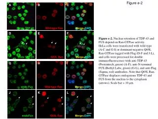
Differentiation of Flag-FOXO3a Localization in WT and p38α-/- MEFs Post Doxorubicin Treatment
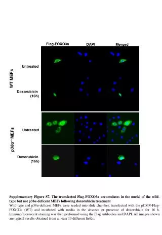
This supplementary figure illustrates the accumulation of transfected Flag-FOXO3a in the nuclei of wild-type MEFs but not in p38α-deficient MEFs after 16 hours of doxorubicin treatment. Both cell types were seeded in slide chambers, transfected with pCMV-Flag-FOXO3a, and treated with doxorubicin. Subsequent immunofluorescent staining using Flag antibodies and DAPI highlighted the differential nuclear localization. The results presented are representative of at least 10 different fields, showcasing the significance of p38α in regulating FOXO3a nuclear translocation under stress conditions.

Differentiation of Flag-FOXO3a Localization in WT and p38α-/- MEFs Post Doxorubicin Treatment
E N D
Presentation Transcript
Flag-FOXO3a DAPI Merged Untreated WT MEFs Doxorubicin (16h) Untreated p38α-/-MEFs Doxorubicin (16h) Supplementary Figure S7. The transfected Flag-FOXO3a accumulates in the nuclei of the wild-type but not p38α-deficent MEFs following doxorubicin treatment Wild-type and p38α-deficent MEFs were seeded into slide chamber, transfected with the pCMV-Flag-FOXO3a (WT) and incubated with media in the absence or presence of doxorubicin for 16 h. Immunofluorescent staining was then performed using the Flag antibodies and DAPI. All images shown are typical results obtained from at least 10 different fields.