Download
1 / 82
820 likes | 834 Vues
Explore the complexities of the nervous system and nervous tissue, including the central and peripheral divisions, sensory input and motor output, and the different types of sensory and motor information. Learn about the structure and function of neurons and the role of support cells in the nervous system.

E N D
THE NERVOUS SYSTEM & NERVOUS TISSUE Human Anatomy Sonya Schuh-Huerta, Ph.D. Leonardo Da Vinci, 1508
Nervous System • Master control & communication system • What makes us uniquely “human” • Great intelligence • Emotions & empathy • Reasoning & problem solving • Strategizing & predicting • Culture (religion, etc.) • …
Nervous System • 3 Overlapping Functions: • Sensory receptors monitor changes inside & outside body • Change a stimulus • Gathered information sensory input • Processes & interprets sensory input • Makes decisions integration • Dictates a response by activating effector organs • Response motor output
Sensory input Integration Motor output Nervous System
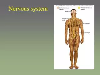
Basic Divisions of Nervous System • Central nervous system (CNS) • Brain & spinal cord • Integrating & command center • Personality traits, emotions, intelligence, etc.
Basic Divisions of Nervous System • Peripheral nervous system (PNS) • Outside the CNS • Consists of nerves extending from brain & spinal cord: • Cranial nerves • Spinal nerves • Peripheral nerves link all regions of body to CNS • Ganglia are clusters of neuronal cell bodies
Brain CNS Spinal cord Nerves PNS Ganglia Basic Divisions of the Nervous System
Sensory Input & Motor Output • Sensory (afferent) signals picked up by sensory receptors • Carried by nerve fibers of PNS to the CNS • Motor (efferent) signals are carried away from the CNS • From brain/spinal cord to organs (muscles, glands)
Sensory Input & Motor Output • Divided according to region they serve: • Somatic body region • Visceral body region • Results in 4 main subdivisions of NS: • Somatic sensory • Visceral sensory • Somatic motor • Visceral motor (=autonomic nervous system)
Types of Sensory & Motor Information Central nervous system (CNS) Peripheral nervous system (PNS) Brain and spinal cord Cranial nerves and spinal nerves Integrative and control centers Communication lines between the CNS and the rest of the body Sensory (afferent) division Motor (efferent) division Somatic and visceral sensory nerve fibers Motor nerve fibers Conducts impulses from the CNS to effectors (muscles and glands) Conducts impulses from receptors to the CNS Somatic sensory fiber Somatic nervous system Autonomic nervous system (ANS) Skin Somatic motor (voluntary) Visceral motor (involuntary) Conducts impulses from the CNS to skeletal muscles Conducts impulses from the CNS to cardiac muscles, smooth muscles, and glands Visceral sensory fiber Stomach Skeletal muscle Motor fiber of somatic nervous system Sympathetic division Parasympathetic division Mobilizes body systems during activity Conserves energy Promotes house- keeping functions during rest Sympathetic motor fiber of ANS Heart Structure Function Sensory (afferent) division of PNS Bladder Parasympathetic motor fiber of ANS Motor (efferent) division of PNS
Basic Divisions of the Nervous System • Somatic sensory • General somatic senses receptors are widely spread • Touch • Pain • Vibration • Pressure • Temperature • (receptors discussed later in Ch 14)
Basic Divisions of the Nervous System • Somatic sensory (cont.) • Proprioceptive senses detect stretch in tendons & muscle • Body sense position & movement of body in space • Special somatic senses (Ch 16) • Hearing • Balance • Vision • Smell & Taste
Basic Divisions of the Nervous System • Visceral sensory • General visceral senses stretch, pain, temperature, nausea, & hunger • Widely felt in digestive & urinary tracts, & reproductive organs • Special visceral senses • Taste & smell often considered special visceral senses
Basic Divisions of the Nervous System • Somatic motor • General somatic motor signals contraction of skeletal muscles • Under our voluntary control! • Often called “voluntary nervous system”
Basic Divisions of the Nervous System • Visceral motor • Regulates the contraction of smooth & cardiac m. • Makes up autonomic nervous system (ANS) • Controls function of visceral organs • Often called “involuntary nervous system” • “Fight or Flight” NS • =Autonomic nervous system (later, Ch 15)
Nervous Tissue • Cells are densely packed & • intertwined • 2 main cell types: • Neurons transmit electrical signals • Support cells (neuroglial or glial cells) • Nonexcitable • Support growth & function of neurons • Surround & wrap neurons
The Neuron • The human body contains billions • of neurons!!! • Basic structural unit of the • Nervous System • Specialized cells that conduct electrical • impulses along their plasma membrane • Nerve impulse (= action potential)
The Neuron • Special characteristics: • 1.) Excitability conduct electrical impulses • 2.) Longevity can live & function for a lifetime! • 3.) Do not divide fetal neurons lose their ability to undergo mitosis; neural stem cells are an exception (olfactory & hippocampal neuron regeneration is an example) • 4.) High metabolic rate require abundant oxygen & glucose! • Neurons die after 5 minutes without oxygen!!!
The Cell Body • Cell body (= soma) • Perikaryon cytoplasm around the nucleus • Size of cell body varies from 5–140 µm • Contains usual organelles plus other structures • Chromatophilic bodies (= Nissl bodies) • Clusters of rough ER & • free ribosomes • Stain darkly • Protein machinery • Renew membranes of • the cell Chromatophilic (Nissl) bodies
The Cell Body • Neurofibrils bundles of intermediate filaments • Form a network between chromatophilic bodies Neurofibril
The Cell Body • Most neuronal cell bodies are: • Located within the CNS! • Protected by bones of skull & • vertebral column • Ganglia (“knot in a string”) • clusters of cell bodies • Lie along nerves in the PNS!
Structure of a Typical Large Neuron Dendrites (receptive regions) Cell body (biosynthetic center and receptive region) Dendrites Neuron cell body Nucleus with nucleolus Neurofibril Nucleus Chromatophilic (Nissl) bodies (b) Nuclei of neuroglial cells Axon (impulse generating and conducting region) Impulse direction Nucleolus Nissl bodies Node of Ranvier Axon terminals (secretory region) Axon hillock Schwann cell (one inter- node) Neurilemma (a) Terminal branches
Neuron Processes • Dendrites • Extensively branch from the cell body • Transmit electrical signals toward the cell body • Chromatophilic bodies only extend into basal part of dendrites & to the base of axon hillock • Function as receptive sites for receiving signals from other neurons
Neuron Processes • Axon • Neuron has only one • Impulse generator & conductor axon hillock • Transmits impulses away from the cell body • Impulses travel to axon terminals • Chromatophilic bodies are absent • No protein synthesis in axon
Neuron Processes • Axons • Neurofilaments, actin • microfilaments, & • microtubules • Provide strength & • structure along • length of axon • Aid in the transport of • substances to & from • the cell body • Axonal transport
Neuron Processes • Axons • Branches along length are infrequent • Axon collaterals • Multiple branches at end of axon • Terminal branches • End in knobs called axon terminals(also called end bulbs or boutons)
Nerve Impulse • Nerve impulse = action potential • Generated at initial segment of the axon (hillock) • Conducted along the axon electrical signal • Causes release of neurotransmitters at axon terminals • Neurotransmitters chemical signals (excite or inhibit neurons) • This is how the neuron • receives & sends signals
Synapses • Site at which neurons communicate • Signals pass across synapse in 1 direction • Presynaptic neuron • Conducts signal toward a synapse • Postsynaptic neuron • Transmits electrical signal away from a synapse
2 Neurons Communicating at a Synapse Presynaptic neuron Axon Axon terminal at synapse Postsynaptic neuron Synapse Dendrite (a) 2 neurons connected by synapses
Types of Synapses • Axodendritic • Between axon terminals of one neuron & dendrites of another • Most common type of synapse! • Axosomatic • Between axons & cell bodies
Synapses • Elaborate cell junctions • Synaptic vesicles in presynaptic neuron • Membrane-bound sacs containing neurotransmitters • Neurotransmitter released & binds to receptor & initiates depolarization of postsynaptic neuron • Mitochondria abundant in axon terminals • Synaptic cleft • Separates the plasma membrane of the 2 neurons
The Synapse – Remember this at NMJ? Nerve impulses Presynaptic axon Microtubule Neurofilament Axon terminal Mitochondrion Vesicle releasing neurotransmitter Synaptic vesicles Synaptic cleft Postsynaptic dendrite (b) Enlarged view of the synapse
Classification of Neurons • Structural classification • Multipolar possess more than 2 processes • Numerous dendrites & one axon • Bipolar possess 2 processes (1 axon, 1 dendrite) • Rare neurons • Found in some special sensory organs • Unipolar (pseudounipolar) possess 1 short, single process
Functional Classification of Neurons • Types of neurons based on functional classification: • Sensory Neurons • Motor Neurons • Interneurons
Functional Classification of Neurons • Sensory (afferent) neurons • Transmit impulses toward the CNS • Virtually all are unipolar neurons • Cell bodies in ganglia outside the CNS • Short, single process divides into: • The central process runs centrally into the CNS • The peripheral process extends peripherally to the receptors
Functional Classification of Neurons • Motor (efferent) neurons • Carry impulses away from the CNS to effector organs • Most Motor neurons are Multipolar • Cell bodies are within the CNS • Form junctions with effector cells • Interneurons (= association neurons) • Most are multipolar • Lie between/connect motor & sensory neurons • Confined to the CNS
Supporting Cells • 6 types of supporting cells: • 4 in the CNS • 2 in the PNS • Provide supportive functions for neurons • Cover nonsynaptic regions of the neurons
Neuroglia in the CNS • Neuroglia • Glial cells have branching processes & a central cell body • Outnumber neurons 10 to 1!!! • Make up half the mass of the brain • Can divide throughout life!!! • Have neuroglial adult stem cells
Neuroglia in the CNS • Astrocytes most abundant glial cell type • Sense when neurons release glutamate • Extract blood sugar from capillaries for energy • Take up & release ions to control environment • around neurons • Involved in synapse formation in developing neural tissue • Produce molecules necessary for neuronal growth • (BDTF) • Propagate calcium signals involved with memory
Neuroglia in the CNS Capillary Neuron Astrocyte (a) Astrocytes are the most abundant CNS neuroglia
Neuroglia in the CNS • Microglia smallest & least abundant glial cell • Phagocytes the macrophages of the CNS • Engulf invading microorganisms & dead neurons • Derived from blood cells called monocytes
Neuroglia in the CNS Neuron Microglial cell (b) Microglial cells are defensive cells in the CNS
Neuroglia in the CNS • Ependymal cells • Line the central cavity of spinal cord & brain • Bear cilia help circulate the cerebrospinal fluid • Oligodendrocytes have few branches • Wrap their cell processes around axons in CNS • Produce myelin sheaths!!!
Neuroglia in the CNS Fluid-filled cavity Ependymal cells Brain or spinal cord tissue (c) Ependymal cells line cerebrospinal fluid–filled cavities. Myelin sheath Process of oligodendrocyte Nerve fibers (d) Oligodendrocytes have processes that form myelin sheaths around CNS nerve fibers.
Neuroglia in the PNS • Satellite cells surround neuron cell bodies within ganglia • Schwann cells surround axons in the PNS • Form myelin sheath around axons of the PNS Satellite cells Cell body of neuron Schwann cells (forming myelin sheath) Nerve fiber Satellite cells and Schwann cells (which form myelin) surround neurons in the PNS.
Myelin Sheaths • Segmented structures composed of the lipoprotein myelin whitish in color • Surround thicker axons • Form an insulating layer • Prevent leakage of electrical current • Increase the speed of impulse conduction! • Like the casing surrounding an electrical wire
Myelin Sheaths in the PNS • Formed by Schwann cells • Develop during fetal period & in the first year of postnatal life • Schwann cells wrap in concentric layers around axon • Cover axon in a tightly packed coil of membranes • Neurilemma • Material external to myelin layers
Myelin Sheaths • Nodes of Ranvier gaps along axon • Gaps in between adjacent Schwann cells • Thick axons are myelinated • Thin axons are unmyelinated • Conduct impulses • more slowly Nodes of Ranvier • What happens at Nodes of Ranvier?