Download
1 / 59
590 likes | 894 Vues
Diseases of the Esophagus. כירורגית חזה מ . ר קפלן. אנטומיה. צינור שרירי באורך 25 ס"מ מחולק ל-3: צוארי, חזי, בטני בתמט במנוחה נמתח עד 2-3 ס"מ בבליעה שטוח ב-2/3 העליונים ומעוגל ב-1/3 התחתון מתחיל מגובה 6 C ועד כ-3 ס"מ מתחת לסרעפת – 11 T האיבר הצר ביותר במערכת העיכול למעט התוספתן. הושט.

E N D
Diseases of the Esophagus כירורגית חזה מ.ר קפלן
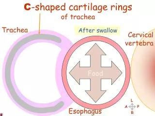
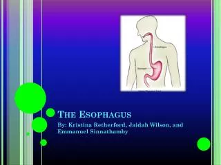
אנטומיה • צינור שרירי באורך 25 ס"מ • מחולק ל-3: צוארי, חזי, בטני • בתמט במנוחה • נמתח עד 2-3 ס"מ בבליעה • שטוח ב-2/3 העליונים ומעוגל ב-1/3 התחתון • מתחיל מגובה 6C ועד כ-3 ס"מ מתחת לסרעפת – 11T • האיבר הצר ביותר במערכת העיכול למעט התוספתן
הושט • 1/3 עליון הוא שריר מפוספס • 1/3 תחתון הוא שריר חלק • 1/3 אמצעי הוא שילוב של השניים • מכיל שני ספינקטרים • מכוסה באפיתל קשקשי
מבנה • לושט 4 שכבות • Mucosa • Submucosa • Muscularis propria • Adventitia • בניגוד לשאר מע' העיכול - אין סרוזה
ספינקטרים • אזורים פונקציונליים ולא אנטומיים • ניתן לזהות ע"י מדידות לחץ • UES: • אזור לחץ גבוה באורך 2-3 ממ בין הפארינקס לושט • סמוך ל- cricopharyngeal muscle
LES: • אזור לחץ גבוה באזור ה-GEJ • בד"כ כ-3 ס"מ מתחת להיאטוס
השכבות השריריות אחראיות לפריסטלטיקה של הושט • שתי שכבות – פנימית צירקולרית וחיצונית אורכית
ניקוז לימפטי • מע' אנסטומוזות נרחבת • מע' ניקוז תת רירית • הכרחי להתפשטות מהירה של גידולים • שליחת גרורות לפני חסימה של החלל • שיעור הישנות גבוה
Surgical Diseases of the Esophagus • Hiatal Hernia • Reflux esophagitis • Esophageal motility disorders • Cancer
Hiatal Hernia • Sliding hiatal hernias are more common – 95% • The lower esophageal sphincter mechanism becomes incompetent • Reflux of acid gastric juice produces a chemical burn • Degree of mucosal injury is a function of the duration of acid contact
Hiatal Hernia • Continued inflammation of the distal esophagus may lead to • mucosal erosion • Ulceration • eventually scarring and stricture • Intestinal metaplasia (Barrett`s esophagus)
Clinical Presentation • Hiatal hernia with reflux is frequently found in patients who are overweight. • Many patients with hiatal hernia have no symptoms. • A burning epigastric or substernal pain or tightness • Usually the pain does not radiate • May be described as a tightness in the chest and can be confused with the pain of myocardial ischemia
Hiatus Hernia - Clinical Presentation • Aggravate • supine or leaning over, alcohol, aspirin, tobacco, and caffeine • Allevaite • Antacid therapy • Late symptoms of dysphagia and vomiting usually suggest stricture formation
Diagnosis- Hiatus Hernia • Anamnesis • Weight loss is a feature due to distal esophageal stricture • Manometry - loss of the lower esophageal high-pressure area • 24-h pH-monitoring – gold standard for GERD • Upper-GI series • Esophagogastric endoscopy - Bx
Treatment – Hiatus Hernia Medical Therapy • Avoidance of gastric stimulants (coffee, tobacco, and alcohol) • Weight loss • Avoid eating several hour before bedtime • PPI
Treatment Hiatus Hernia - Surgical • Failure of PPI treatment • Correct the anatomic defect • Prevent the reflux of gastric
Hiatus Hernia • Complications post surgery • 3-10% • inability to belch or vomit- the "gas-bloat" syndrome • Vagal injury • The wrap itself • Dysphagia • Disruption of the repair with recurrent symptoms – 5% • esophageal perforation • Splenic injury • Penumothorax
Esophageal Motility DisordersAchalasia • Failure of the high-pressure zone sphincter to relax • Not due to spasm • Painless dysphagia • Progressive dilation of the proximal esophagus
Esophageal Motility DisordersAchalasia -- Clinical Presentation • Dysphagia • Regurgitation of undigested food • Weight loss • Pain is uncommon • Aspiration pneumonia is common • Complain of spitting up foul-smelling secretions when simply leaning forward
Esophageal Motility DisordersAchalasia -- Diagnosis • Generally first confirmed by contrast studies of the esophagus • Dilation of the proximal esophagus is classic • Esophageal diverticula may be present at any level • Esophageal manometry – gold standard
Esophageal Motility DisordersAchalasia -- Treatment • Medical treatment has generally not been helpful • Invasive endoscopic procedure • forceful dilation – effective - 60%. 4% perforations • Botoxtm – 50% recurrence within 6 months • Surgical • Transaction of muscle - Heller myotomy • Esophagectomy – sigmoid esophagus, failure of more than one myotomy, or an undilatable stricture
Esophageal Motility DisordersEsophageal Diverticulum • The second most common manifestation of esophageal motility disorders • Pulsion orTraction, depending on the mechanism that leads to their development
Esophageal Motility DisordersEsophageal Diverticulum • Upper third cervical esophageal diverticula - usually pulsion • Cervical diverticula, or Zenker's • pulsion • related to dysfunction of the cricopharyngeal muscle • complain of regurgitation of recently swallowed food • putrid breath odor
Treat the underlying condition! • Excision of the diverticula • Myotomy of the cricopharyngeal muscle
Esophageal Motility DisordersEsophageal Diverticulum – Zenker’s
Esophageal Motility DisordersEsophageal Diverticulum • Middle-third esophageal diverticula • Traction • Not related to esophageal dysmotility • Result of mediastinal inflammation • Usually asymptomatic and do not warrant treatment.
Esophageal Motility DisordersEsophageal Diverticulum • Diverticula of the distal third of the esophagus • Dysfunction of the esophagogastric junction • Stricture • antireflux surgical procedures • Achalasia • Excision of the diverticula • Always - correction of the underlying pathologic process
Esophageal NeoplasmsBenign • Exceedingly rare – in middle and distal 1/3 • Leiomyomas are the most common intramural tumors 1) potential for malignant degeneration appears to be quite low 2) indent the lumen of the esophagus on contrast radiography 3) tend to grow progressively and cause dysphagia 3) Excised for possible dysphagia and malignancy
Barrett`s esophagus • GERD - Chronic acid reflux • Columnar intestinal metaplasia • 40 times risk for AdenoCA • May progress to • Low grade dysplasia • High grade dysplasia (stage 0 carcinoma) • Invasive AdenoCA
Esophageal NeoplasmsMalignant • 30% are squamous cell carcinomas • 70% are Adenocarcinomas • < 1% are • malignant melanoma • Adenoid cystic tumors • Sarcomas
Esophageal NeoplasmsMalignant • Origin • SCC – squamous epithelium • AdenoCA – Barrett`s esophagus • Commonly occurs in association with alcohol and/or tobacco abuse • Etiology has been related to diet, vitamin deficiency, poor oral hygiene, surgical procedures, and a number of premalignant conditions, (caustic burns, Barrett's esophagus, radiation, esophageal diverticula).
Esophageal NeoplasmsMalignant • Vague symptoms at early stages • Weight loss and pain may be present • Dysphagia – obstruction of >30% • Odynophagia • Iron deficiency anemia
Esophageal NeoplasmsMalignant -- Diagnosis • Endoscopy and biopsy of the lesion • Endoscopic US – T & N staging • Chest & Abdominal CT – M staging • PET CT
TNM Staging • T1 lamina propria or submucosa • T1a muscularis mucosae • T1b submucosa • T2 - muscularis propria • T3 - adventitia • T4 - adjacent structures • N0 - No regional lymph node metastasis • N1 - 1 to 2 regional lymph nodes • N2 - 3 to 6 • N3 - >6
Esophageal NeoplasmsMalignant -- Treatment • Early disease – stage I,(IIa ?) • Surgery • Locally advanced disease – stage IIb, III • Neoadjuvant therapy • Surgery
Esophageal NeoplasmsMalignant -- Treatment • Surgical techniques • Transhiatal esophagectomy • Transthoracic esophagectomy • En-block esophagectomy
טיפול כירורגי טיפול כירורגי למטרת ריפוי טיפול כירורגי כפליאציה? כירורגיה- Gold standard
ניתוחים- (לפי מיקום) • ושט צווארי - three field esophagectomy )א.א.ג+ חזה+ כירורגיה) jejunal flap/ stomach • ושט חזי - three field esophagectomy Ivor Lewis/ lt thoracotomy • ושט תחתון - Ivor Lewis או Trans Hiatal • "צומת ושט קיבה" - Ivor Lewis או lt thoracotomy במשלב עם כריתת קיבה
טיפול כירורגי טכניקה כירורגית • כניסה? • כמה ושט לכרות? כמה בלוטות להסיר? • "תחליף ושט"? טכניקת השקה? מאפייני מטופל • מיקום השאת? • מאפייני מטופל? • ניסיון כירורגי?
Trans Hiatal Approach • גישה נפוצה • 2 חתכים • OPEN/ LAP
Trans Hiatal Approach • יתרונות • השקה בצוואר • תחלואה • חסרונות • אונקולוגיה
Ivor Lewis approach • יתרונות • אונקולוגיה • תחליף ושט • חסרונות • תחלואה, תמותה.