Pacemaker Patient Follow-up Module 8
Pacemaker Patient Follow-up Module 8. Objectives. List the steps for performing a pacemaker follow-up Locate threshold, sensing and impedance data on a Quick Look™ screen Identify broad trends in Cardiac Compass ® and HF Management Reports


Pacemaker Patient Follow-up Module 8
E N D
Presentation Transcript
Objectives • List the steps for performing a pacemaker follow-up • Locate threshold, sensing and impedance data on a Quick Look™ screen • Identify broad trends in Cardiac Compass® and HF Management Reports • Identify additional resources or information useful for performing patient follow-up
Pacemaker Patient Follow-up Routine device follow-up • Typically 1 per year for chronic single chamber and 2 per year for chronic dual chamber pacemakers • These guidelines evolved because the primary purpose of the follow-up was to evaluate the device • Increasing sophistication of follow-up tools and the diagnostics the devices provide, may be changing the purpose of the follow-up, but have not affected the guidelines
Pacemaker Patient Follow-up Steps Evaluating the device • Determine the battery voltage • Check the lead impedance • Test capture thresholds • Test sensing thresholds • Perform a magnet/non-magnet test (required by Medicare in some US states)
Pacemaker Patient Follow-upEvaluating the Device • Battery voltage • To verify the pacemaker’s ability to operate and estimate the remaining longevity • Lead impedance • To verify that the leads are electrically intact • Capture thresholds • To verify an appropriate pacing safety margin • Sensing thresholds • To verify an appropriate sensing safety margin • To observe the underlying rhythm • Magnet/non-magnet test • To verify Recommended Replacement Time (RRT) has not been reached
Battery voltage Estimate of remaining time to replacement Most modern devices provide this estimate in some fashion Most modern devices will also indicate impending elective or recommended replacement Lead impedance Measured with interrogation Long-term trends, if available, are very useful Pacing thresholds In some devices this test is automatic and performed routinely (e.g., daily) Sensing thresholds In some devices this test is automatic and performed routinely (e.g., every intrinsic event) Magnet test Formerly only way to determine if Recommended Replacement Time (RRT) had been reached Less of a need as devices now routinely report RRT condition Pacemaker Evolution for the Follow-up Clinic
Battery VoltageEstimating Remaining Longevity • Max voltage in a pacemaker battery is typically 2.8 V • RRT at 2.5 Volts • Voltage information is provided on the programmer • Many devices estimate remaining longevity based on % pacing, lead impedances, and pacing outputs • Magnet Test evolved as a way to evaluate if RRT criteria is met via a simple telephone or in-clinic check • When a magnet is applied, verify if the pacemaker shows normal magnet behavior or RRT magnet behavior • For some pacemakers, a magnet initiates a Threshold Margin Test (TMT)
Resource Pacemaker And ICD Encyclopedia • All device manufactures share essential device information • All publish encyclopedias • Medtronic’s version is available on-line, as a download for both Palm and Pocket PC • All include information on leads, devices, model numbers, x-ray identification, etc. • Includes Beginning-of-Life (BOL) magnet mode and rates • RRT magnet mode and rates • Other RRT indicators (e.g., pulse width “stretching”)
Status Check • Determine the magnet and non-magnet modes, and rates for a Kappa® 700, Model KDR731. Click for answer
Status Check • Find the Estimated Longevity Click for answer Battery Voltage can be found by selecting “Battery and Lead Measurements” under the Data icon.
Magnet Testing • If required: • Use a donut magnet (or the programming head) • Record the ECG with and without the magnet • Medicare guidelines call for a 30-second recording of each • Observe the ECG for asynchronous pacing with the magnet • Measure the pacing rate • Some modern devices provide a specific magnet test as an option on their programmer
Lead Impedance • Can you recall why knowing the lead impedance is important? • Because the lead impedance can warn you of a lead insulation failure or a lead conductor fracture • At implant, it could indicate a loose set screw • Can you recall the expected range of lead impedances? • Normally, 300-1000 Ω, although some specially designed leads may be higher Click for answer Click for answer
These trends show stable impedances. For more information, touch the chevron. Status Check • Find the lead impedances Click for answer
Pacing Thresholds • Can you recall the pacing output safety margin on a chronic system? • Multiply the amplitude threshold by two • Can you recall the maximum you would want to program the amplitude, assuming a threshold of < 1.0 V? • Normally, try to keep the output amplitude at < 2.5 V • Patient safety is paramount Click for answer Click for answer
These trends show stable thresholds. Note how the atrial threshold has decreased since implant. For more information, touch the chevron. Status Check • Find the pacing thresholds Click for answer
Threshold TestingManual Methods • The device automatically performs threshold tests But where would you find a manual threshold (or other) test? Click for answer
Status Check • Recall the definition of the pacing threshold… • The minimum output at which the myocardium is consistently captured, outside of its refractory period. Click for answer
Performing a Manual Pacing Threshold Test • Process • Tell the patient what you are going to do • Force pacing • Periodically lower the test value (amplitude or pulse width) • Stop the test once Loss-of-Capture (LOC) is seen on the ECG • The value just above LOC is the threshold 0.75 V 1.25 V 1.00 V
To see the P- and R-wave trends, touch the chevron. Status Check • Find the P- and R-wave measurements Click for answer
Sensing TestsManual Methods • This device automatically measures P- and R-waves But where would you find a manual sensing (or other) test? Click for answer
Status Check • What is the definition of the sensing threshold? • The minimum signal size required to inhibit (or trigger) the pacemaker Click for answer
Performing a Manual Sensing Test • Process • Tell the patient what you are going to do • Reduce (if necessary) the pacing rate to allow the intrinsic events to occur • Periodically increase the test value (e.g., for 0.5 mV to 1.0 mV) • Observe the ECG for loss of inhibition (pacing despite the presence of P- or R-waves) • The value, just lower than when pacing occurs, is the size of the P- or R-wave
Routine Testing • With almost all manufacturers, routine threshold and sensing tests can be performed on a programmer interface that guides you through the steps • Observe the patient carefully for symptoms or complaints • Follow the on-screen directions and closely observe the ECG for changes
Other Routine Tests • Underlying rhythm • Can frequently ascertain the underlying rhythm during the sensing test • Optionally, you can perform this test with the programmer • Retrograde conduction • Etc. • Done per clinic protocol, or only if needed
So We’ve Checked the Pacemaker • Is that all there is to it? • Well… • Is Rate Response programmed appropriately? • Can the patient achieve rates to support activities of daily living? • Is the patient having any arrhythmias? • What kind of arrhythmias, what rates, what triggers? • Can we program the device to extend its longevity? • Can we encourage intrinsic events? …are just some of the questions we can answer by looking at the device diagnostics.
Always evaluate the rate histograms Look for a “stair-case” distribution of rates Ask about the patient’s ability to achieve their desired levels of activity Optimizing Pacemakers for Patients Rates below the LRL may occur because of a timing anomaly. Rates above the UTR/USR may indicate an arrhythmia.
Optimizing Pacemakers for Patients • Always evaluate for the presence of arrhythmia • What type? • Is the patient symptomatic? Cardiac Compass is a simple way to stay informed of trends in the patient’s atrial arrhythmia burden. Cardiac Compass also provides trends on the ventricular response to these arrhythmia.
Episode Specific Diagnostics In pacemakers, stored EGMs can be useful: • Confirming accuracy of other arrhythmia diagnostics • For example – is oversensing triggering arrhythmia collection? • Collecting arrhythmia triggers • An EGM is an ECG recorded via the pacemaker’s leads and stored in the device Stored EGM of an episode of AF
Optimizing Pacemakers for Patients • Always evaluate the percent pacing • Many devices have a percent pacing counter, or look to the histograms for this information • Device longevity is affected by pacing outputs • High outputs decrease longevity • Low outputs (below about 2.0 V) don’t have as dramatic of an affect • So reducing the percentage of unnecessary pacing will improve device longevity and there may be other benefits to patients (more later)
Reducing the Pacing Percentage Managing AV delays • Use auto AV extension algorithms (e.g., Medtronic’s Search AV+) with Auto-PVARP options to: • Reduce unnecessary right ventricular pacing • Allow higher UTR • Guard against retrograde conduction
Reducing the Pacing Percentage Search AV+ example: • AV intervals are scanned • If the majority end in pacing (8/16) the AV delay is automatically increased • Note: AV scanning algorithms will typically still result in ventricular pacing about 40-50% of the time.
Using Other Diagnostics • Some devices include indicators of HF status and can be very useful • For example: HR variability, Average day/night rates, Activity trends • Otherwise be guided by: • The patient’s diagnosis, complaints, symptoms
Clinic EvolutionTaking Advantage of Pacemaker Automaticity • Devices do not routinely require extensive testing to confirm they are operating correctly • But when troubleshooting a particular problem is required, most devices today offer detailed data that may help assist with analysis of device, lead and disease progression concerns • If all tests can be performed automatically, and if we could view all device diagnostics remotely, why not: • Use the pacemaker clinic only when a device has a problem or requires reprogramming • Allow the physician to determine if routine clinic visits are warranted?
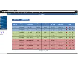
Medtronic CareLink® Network Example Clinic A ****** Click to Login
CareLink™ Network Example Pick the patient transmission Note: the patient information is fictitious
A-A interval 705ms V-V interval 705ms Status CheckCase 1 This patient has had a DDDR pacemaker for 6 years • Functioning normally • On a routine telephone check, the following strip is transmitted • The patient’s magnet is in place • What conclusions can you draw? Click for answer Asynchronous pacing Normal magnet behavior, operating in DOO mode at 85 ppm
Status CheckCase 2 This patient was implanted about 6 months ago for SND • He came to the clinic for a wound check 2 weeks post op. This is first IPG clinic visit. • This (hypothetical) pacemaker requires that you do all the testing. • How is the device currently programmed? Click for answer
Status CheckCase 2 Results of pacemaker testing: • Battery voltage: 2.78 V • Sensing: • P-waves 2.0-2.5 mV • R-waves >22.8 mV • Underlying rhythm Sinus Brady at 50 bpm • Thresholds • Atrial 1.0 V tested at 0.4 ms • Ventricular 0.8 V tested at 0.4 ms
Status CheckCase 2 • What programming changes (if any) would you recommend? • Re-program the A and V outputs to 2.0 V at 0.4 ms is reasonable. The leads should be stable at 6 months post implant. • Would you run any other tests or investigate any further? • The underlying rhythm indicates intact conduction. Consider programming a Search AV algorithm to reduce unncessary ventricular pacing. • Check the rate histograms and question the patient for the adequacy of his rate response. • Check other diagnostics for the presence of arrhythmia. Click for answer Click for answer
Status CheckCase 3 • This elderly patient had a DDDR implanted for sinus node dysfunction 8 weeks ago • Discovered sinus node disease with pauses on routine physical • Past medical history includes hypertension • She denies palpitations • Treatment: Hydrochlorothiazide (blood pressure medication) • Patient comes to the clinic for her first routine check • All values within normal and expected limits • Atrial and Ventricular outputs reduced to a 2.5x safety margin • No other changes made
Status CheckCase 3 Cardiac Compass Report Is she having AF? How much AF is she having? Click for answer Episodes totaling 8 hours on one day, about 4 hours on two days, and several shorter episodes 1 min to 1 hour durations thereafter.
Status CheckCase 3 Cardiac Compass Report Is the ventricular response to AF well controlled? Click for answer Yes, as a very low percent of the ventricular response to AF is > 100 bpm.
A Couple of Questions • In patients with paroxysmal AF (PAF), how likely are they to experience symptoms of AF? • What is the risk of stroke or CVA to someone with untreated AF? Click for answer Asymptomatic PAF occurs 12.1 times as often as symptomatic PAF in symptomatic patients [Page et al, Circulation 89(1):224-7, 1994] Asymptomatic PAF occurs in 22-27% of patients with clinical improvement [Wolk et al, Int J Cardiol 54:207-211, 1996] Click for answer 5-7% per year. The take-away: AF is silent and dangerous.
Status CheckCase 3 • Based on the Cardiac Compass® data, the MD decides to add Sotalol 160 bid and Coumadin • The patient returns 6 months later • Is her arrhythmia status improved? Click for answer Yes, it has improved. Her last episode was at the end of May.
A Couple More Questions • For this patient, how would you make the diagnosis of PAF without these kinds of diagnostics? • She had no symptoms • She was not in AF on clinic visits • For this patient, how would you evaluate the effectiveness of your treatment without these kind of diagnostics? • Her Medication regimen was not that effective in suppressing her AF • She had no symptoms • She was not in AF on clinic visits
Brief Statements Indications • Implantable Pulse Generators (IPGs) are indicated for rate adaptive pacing in patients who ay benefit from increased pacing rates concurrent with increases in activity and increases in activity and/or minute ventilation. Pacemakers are also indicated for dual chamber and atrial tracking modes in patients who may benefit from maintenance of AV synchrony. Dual chamber modes are specifically indicated for treatment of conduction disorders that require restoration of both rate and AV synchrony, which include various degrees of AV block to maintain the atrial contribution to cardiac output and VVI intolerance (e.g. pacemaker syndrome) in the presence of persistent sinus rhythm. • Implantable cardioverter defibrillators (ICDs) are indicated for ventricular antitachycardia pacing and ventricular defibrillation for automated treatment of life-threatening ventricular arrhythmias. • Cardiac Resynchronization Therapy (CRT) ICDs are indicated for ventricular antitachycardia pacing and ventricular defibrillation for automated treatment of life-threatening ventricular arrhythmias and for the reduction of the symptoms of moderate to severe heart failure (NYHA Functional Class III or IV) in those patients who remain symptomatic despite stable, optimal medical therapy and have a left ventricular ejection fraction less than or equal to 35% and a QRS duration of ≥130 ms. • CRT IPGs are indicated for the reduction of the symptoms of moderate to severe heart failure (NYHA Functional Class III or IV) in those patients who remain symptomatic despite stable, optimal medical therapy, and have a left ventricular ejection fraction less than or equal to 35% and a QRS duration of ≥130 ms. Contraindications • IPGs and CRT IPGs are contraindicated for dual chamber atrial pacing in patients with chronic refractory atrial tachyarrhythmias; asynchronous pacing in the presence (or likelihood) of competitive paced and intrinsic rhythms; unipolar pacing for patients with an implanted cardioverter defibrillator because it may cause unwanted delivery or inhibition of ICD therapy; and certain IPGs are contraindicated for use with epicardial leads and with abdominal implantation. • ICDs and CRT ICDs are contraindicated in patients whose ventricular tachyarrhythmias may have transient or reversible causes, patients with incessant VT or VF, and for patients who have a unipolar pacemaker. ICDs are also contraindicated for patients whose primary disorder is bradyarrhythmia.
Brief Statements (continued) Warnings/Precautions • Changes in a patient’s disease and/or medications may alter the efficacy of the device’s programmed parameters. Patients should avoid sources of magnetic and electromagnetic radiation to avoid possible underdetection, inappropriate sensing and/or therapy delivery, tissue damage, induction of an arrhythmia, device electrical reset or device damage. Do not place transthoracic defibrillation paddles directly over the device. Additionally, for CRT ICDs and CRT IPGs, certain programming and device operations may not provide cardiac resynchronization. Also for CRT IPGs, Elective Replacement Indicator (ERI) results in the device switching to VVI pacing at 65 ppm. In this mode, patients may experience loss of cardiac resynchronization therapy and / or loss of AV synchrony. For this reason, the device should be replaced prior to ERI being set. Potential complications • Potential complications include, but are not limited to, rejection phenomena, erosion through the skin, muscle or nerve stimulation, oversensing, failure to detect and/or terminate arrhythmia episodes, and surgical complications such as hematoma, infection, inflammation, and thrombosis. An additional complication for ICDs and CRT ICDs is the acceleration of ventricular tachycardia. • See the device manual for detailed information regarding the implant procedure, indications, contraindications, warnings, precautions, and potential complications/adverse events. For further information, please call Medtronic at 1-800-328-2518 and/or consult Medtronic’s website at www.medtronic.com. Caution: Federal law (USA) restricts these devices to sale by or on the order of a physician.
Brief Statement: Medtronic Leads Indications • Medtronic leads are used as part of a cardiac rhythm disease management system. Leads are intended for pacing and sensing and/or defibrillation. Defibrillation leads have application for patients for whom implantable cardioverter defibrillation is indicated Contraindications • Medtronic leads are contraindicated for the following: • ventricular use in patients with tricuspid valvular disease or a tricuspid mechanical heart valve. • patients for whom a single dose of 1.0 mg of dexamethasone sodium phosphate or dexamethasone acetate may be contraindicated. (includes all leads which contain these steroids) • Epicardial leads should not be used on patients with a heavily infracted or fibrotic myocardium. • The SelectSecure Model 3830 Lead is also contraindicated for the following: • patients for whom a single dose of 40.µg of beclomethasone dipropionate may be contraindicated. • patients with obstructed or inadequate vasculature for intravenous catheterization.
Brief Statement: Medtronic Leads (continued) Warnings/Precautions • People with metal implants such as pacemakers, implantable cardioverter defibrillators (ICDs), and accompanying leads should not receive diathermy treatment. The interaction between the implant and diathermy can cause tissue damage, fibrillation, or damage to the device components, which could result in serious injury, loss of therapy, or the need to reprogram or replace the device. • For the SelectSecure Model 3830 lead, total patient exposure to beclomethasone 17,21-dipropionate should be considered when implanting multiple leads. No drug interactions with inhaled beclomethasone 17,21-dipropionate have been described. Drug interactions of beclomethasone 17,21-dipropionate with the Model 3830 lead have not been studied. Potential Complications • Potential complications include, but are not limited to, valve damage, fibrillation and other arrhythmias, thrombosis, thrombotic and air embolism, cardiac perforation, heart wall rupture, cardiac tamponade, muscle or nerve stimulation, pericardial rub, infection, myocardial irritability, and pneumothorax. Other potential complications related to the lead may include lead dislodgement, lead conductor fracture, insulation failure, threshold elevation or exit block. • See specific device manual for detailed information regarding the implant procedure, indications, contraindications, warnings, precautions, and potential complications/adverse events. For further information, please call Medtronic at 1-800-328-2518 and/or consult Medtronic’s website at www.medtronic.com. Caution: Federal law (USA) restricts this device to sale by or on the order of a physician.