Pacemaker Automatic Features Module 10
Pacemaker Automatic Features Module 10. Topics. Atrial and Ventricular Capture Management ® Sensing Assurance Auto Adjusting Sensitivity Lead Monitor and Polarity Switch Rate Response. Capture Management ® Objectives. Describe the value of Atrial and Ventricular Capture Management ®

Pacemaker Automatic Features Module 10
E N D
Presentation Transcript
Topics • Atrial and Ventricular Capture Management® • Sensing Assurance • Auto Adjusting Sensitivity • Lead Monitor and Polarity Switch • Rate Response
Capture Management® Objectives • Describe the value of Atrial and Ventricular Capture Management® • Recall the basic operation of ACM and VCM • Identify how to program ACM and VCM
Capture Management® • Why is it important? • Patient Safety • Device Longevity • Troubleshooting Information • How is it conducted? • Automatic algorithms that mimic what a person would do in a clinic
Assess Patient’s Rhythm Determine Threshold Program Appropriately Capture Management® - Three Categories
Assess Patient’s Rhythm Determine Threshold Program Appropriately Capture Management®
Assess Patient’s Rhythm Determine Threshold Program Appropriately Capture Management® • When you get ready to run a threshold test manually: • What questions do you ask? • Why do you ask these questions? Would you run a ventricular threshold test on a patient whose underlying rhythm is AF with a ventricular response of 120 bpm? NO
Assess Patient’s Rhythm Determine Threshold Program Appropriately Capture Management® • VCM Stability check • Are conditions favorable to conduct a search? • What is the rate? (typically lower than 90 – 100 bpm) • What is rhythm? (defined by few VR/AR/VSP/PVC) • Are there feature interaction? (No RDR or Mode Switch in progress) • If conditions are unfavorable, what do you think happens? • The threshold test is postponed • If conditions are favorable • Move on to determining the threshold
Assess Patient’s Rhythm Determine Threshold Program Appropriately Capture Management®
Assess Patient’s Rhythm Determine Threshold Program Appropriately Capture Management® • Decide how to pace • Why do you need to decide how to pace? • To force pacing • What are your options for forcing pacing? • Increase the rate in a non-tracking mode (VVI/R, DDI/R) • How much? • Decrease the AV delay in a tracking mode (DDD/R) • How much?
Assess Patient’s Rhythm Determine Threshold Program Appropriately Capture Management® • Choose the type of test • The device runs a strength duration test for Ventricular Capture Management 1.50 V Pulse Width Threshold 1.25 V 1.0 V 0.75 V Amplitude Threshold 0.5 V 0.4 ms 1.0 ms
Assess Patient’s Rhythm Determine Threshold Program Appropriately Capture Management® • Test paces, backup paces, and support cycles • Test pace- pace delivered to determine capture • Backup pace- pace delivered immediately following each test pace to ensure there are no dropped beats Q: If you start running a threshold test on a pacer dependent patient, and immediately see Loss of Capture, what do you do? A: Stop the test
Assess Patient’s Rhythm Determine Threshold Program Appropriately Capture Management® • Test paces, backup paces, and support paces • Support cycles- a series of three events (paced or sensed) that allow for the heart to function as it would without being tested Q: As you are running a threshold test on a patient at an accelerated rate to force pacing, the patient becomes extremely symptomatic, what do you do? A: Stop the test
Ventricular Capture Management® Test & Back-up Pace Test & Back-up Pace Support Events
Assess Patient’s Rhythm Determine Threshold Program Appropriately Capture Management® Threshold Search • Starting Output • Start at an output that capture should occur • Capture Management® starts its threshold search at the previous measured threshold value Starting Output Decrement Output When you are performing a threshold, how do you shorten the test? Ending Output
Assess Patient’s Rhythm Determine Threshold Program Appropriately Capture Management® Threshold Search • Decrement Output • Lower output (amplitude or pulse width) one step at a time • Ventricular Capture Management® performs Strength Duration Test • Tests amplitude at 0.4 ms PW • Tests PW at 2X amplitude threshold • Atrial Capture Management® performs an Amplitude Decrement Test Starting Output Decrement Output Ending Output
Assess Patient’s Rhythm Determine Threshold Program Appropriately Capture Management® Threshold Search • Ending Output • Capture Management® confirms capture Starting Output What do you do when you see Loss of Capture (LOC)? Decrement Output Programmer Stop the test Analyzer Increase output to confirm capture Ending Output
Capture Management® • Identify Loss of Ventricular Capture How do you identify Loss of Capture (LOC) when running a ventricular pacing threshold? VVI DDD vs.
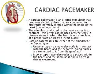
Evoked Response Sensing • All ventricular capture verification algorithms: • Rely on sensing the evoked response to the test pace • The characteristics of this response is how the pacemaker determines if the myocardium is captured • If the characteristics of the evoked response signal differ from what is expected, the pacemaker assumes LOC • Evoked response sensing can be affected by • Lead tissue interface (acute vs. chronic lead) • Lead Polarization • Tip-to-Ring spacing • Lead tip design • Other factors
Capture Management® • Identify Loss of Atrial Capture How do you identify capture when you run an atrial pacing threshold?
Capture Management® • Atrial Chamber Reset Method • Device makes sure that there are no intrinsic atrial events competing with the pacing rate Test AP No Atrial Sensein the AV Interval (Capture)
Capture Management® • AV Conduction Method • Device looks for a conducted R-wave at the predicted time after an atrial pace AP-VS interval following test pace is monitored; if timing is consistent with support pace “expected” window, device records as capture. AP-VS events that result from back-up paces (LOC) will lag by approximately 70 ms from the “expected” window of a test pace.
Assess Patient’s Rhythm Determine Threshold Program Appropriately Capture Management®
Assess Patient’s Rhythm Determine Threshold Program Appropriately Capture Management® • Adapts the output downward in one step decrements (0.125V) • Never below the programmable Minimum Amplitude (Nominal = 2.5V) • Applies the programmable safety margin (Nominal = 2X) to the amplitude at 0.4 ms pulse width • Adapts upward, as needed, to maintain the safety margin • Acute Phase • Output may rise • Will not adapt downward until the Acute Phase expires (N=112 days)
Advanced Pacemaker OperationsTools to Manage Pacemaker Sensing
Sensitivity Management Objectives • State a reason why fixed sensing safety margins may not be adequate • Identify three clinical areas affected by inappropriate sensing • Recall two of the mechanisms for managing sensing automatically
Sensitivity Management • Programming a fixed sensing safety margin may not accommodate for these and other potential changes What factors can change a patient’s P- and R-wave amplitudes? Antiarrhythmia Medications Lead Maturation Atrial Arrhythmias Exercise Ventricular Arrhythmias Myocardial Infarction
Sensitivity Management Normal Operation Therapy Diagnostics
Atrial Bipolar High 2.8 mV 5.6xSensitivity Adequate 2.0 mV 4xSensitivity Low Current Sensitivity 0.5 mV Sensitivity Management • Sensing Assurance • Adapts Sensitivity based on target safety margins, to automatically provide safe sensing margins
Sensitivity Management • Auto Adjusting Sensitivity • Adjusts the sensitivity fence on a beat-by-beat basis
Rate Response Objectives • State the clinical reason for rate response pacing • Recall why rate response pacing works • List the implantations industry uses for rate response pacing
Rate Responsive Pacing • Introduced in the mid-80’s by Medtronic • Why was it one of the most significant developments in pacing? • When one exercises, metabolic demand increases. To meet this demand, cardiac output needs to increase. • What contribution does increasing heart rate make to increasing the cardiac output? Click for Answer Up to a 500% increase over the resting cardiac output. No other component of cardiac output has this significant of a contribution.
Rate Responsive Pacing • This is designated by the “R” in DDDR, AAIR, or VVIR… • It is accomplished by using a sensor to indicate changing metabolic demand • The sensor then modifies the pacing rate • Think of it as a dynamic lower rate or dynamic escape interval • DDDR 60 -130 means: • The heart will not be paced at rates below 60 bpm • The heart may be paced at rates between 60 -130 bpm, based on the information from the rate response sensor • The heart will not be paced at rates above 130 bpm
Rate Response Sensors Many types have been developed with various advantages and disadvantages
Rate Response Sensors • In use today • Accelerometer • Piezo-electric crystal • Combination of MV + Accelerometer or MV + P-E crystal • Combination of QT + Piezo-electric crystal
Rate Response • Rate responsive (also called rate modulated) pacemakers provide patients with the ability to vary heart rate when the sinus node cannot provide the appropriate rate • Rate responsive pacing is for patients who may benefit from increased pacing rates concurrent with increases in activity, such as: • Patients who are chronotropically incompetent (heart rate cannot reach appropriate levels during exercise, or meet other metabolic demands) • Patients in chronic atrial fibrillation with too slow of a ventricular response to meet metabolic demands
Rate Responsive Pacing • Cardiac output (CO) is determined by the combination of stroke volume (SV) and heart rate (HR) • SV X HR = CO • Changes in cardiac output depend on the ability of the HR and SV to respond to metabolic requirements
Rate Responsive Pacing • SV reserves can account for increases in cardiac output of up to 50% • HR reserves can nearly triple total cardiac output in response to metabolic demands
Rate Responsive Pacing • When the need for oxygenated blood increases, the pacemaker ensures that the heart rate increases to provide additional cardiac output Adjusting Heart Rate to Activity Normal Heart Rate Rate Responsive Pacing Fixed-Rate Pacing Daily Activities
A Variety of Rate Response Sensors Exist • Those most accepted in the market place are: • Activity sensors that detect physical movement and increase the rate according to the level of activity • Minute ventilation sensors measure the change in respiration rate and tidal volume via transthoracic impedance readings
Activity sensors employ a piezoelectric crystal that detects mechanical signals produced by movement The crystal translates the mechanical signals into electrical signals that in turn increase the rate of the pacemaker Rate Responsive Pacing Piezoelectric crystal
Rate Responsive Pacing • Minute Ventilation (MV) is the volume of air introduced into the lungs per unit of time • MV has two components: • Tidal volume - the volume of air introduced into the lungs in a single respiration cycle • Respiration rate - the number of respiration cycles per minute
Rate Responsive Pacing • Minute ventilation can be measured by calculating the changes in electrical impedance across the chest cavity to calculate changes in lung volume over time
Status Check • Evaluate this rhythm strip • IPG is programmed to 60-130bpm • What are the atrial and ventricular rates? • What operation is in effect? Click for Answer • A and V rates are about 79 bpm • Rate responsive pacing in the atrium with intrinsic AV conduction
Brief Statements Indications • Implantable Pulse Generators (IPGs) are indicated for rate adaptive pacing in patients who ay benefit from increased pacing rates concurrent with increases in activity and increases in activity and/or minute ventilation. Pacemakers are also indicated for dual chamber and atrial tracking modes in patients who may benefit from maintenance of AV synchrony. Dual chamber modes are specifically indicated for treatment of conduction disorders that require restoration of both rate and AV synchrony, which include various degrees of AV block to maintain the atrial contribution to cardiac output and VVI intolerance (e.g. pacemaker syndrome) in the presence of persistent sinus rhythm. • Implantable cardioverter defibrillators (ICDs) are indicated for ventricular antitachycardia pacing and ventricular defibrillation for automated treatment of life-threatening ventricular arrhythmias. • Cardiac Resynchronization Therapy (CRT) ICDs are indicated for ventricular antitachycardia pacing and ventricular defibrillation for automated treatment of life-threatening ventricular arrhythmias and for the reduction of the symptoms of moderate to severe heart failure (NYHA Functional Class III or IV) in those patients who remain symptomatic despite stable, optimal medical therapy and have a left ventricular ejection fraction less than or equal to 35% and a QRS duration of ≥130 ms. • CRT IPGs are indicated for the reduction of the symptoms of moderate to severe heart failure (NYHA Functional Class III or IV) in those patients who remain symptomatic despite stable, optimal medical therapy, and have a left ventricular ejection fraction less than or equal to 35% and a QRS duration of ≥130 ms. Contraindications • IPGs and CRT IPGs are contraindicated for dual chamber atrial pacing in patients with chronic refractory atrial tachyarrhythmias; asynchronous pacing in the presence (or likelihood) of competitive paced and intrinsic rhythms; unipolar pacing for patients with an implanted cardioverter defibrillator because it may cause unwanted delivery or inhibition of ICD therapy; and certain IPGs are contraindicated for use with epicardial leads and with abdominal implantation. • ICDs and CRT ICDs are contraindicated in patients whose ventricular tachyarrhythmias may have transient or reversible causes, patients with incessant VT or VF, and for patients who have a unipolar pacemaker. ICDs are also contraindicated for patients whose primary disorder is bradyarrhythmia.
Brief Statements (continued) Warnings/Precautions • Changes in a patient’s disease and/or medications may alter the efficacy of the device’s programmed parameters. Patients should avoid sources of magnetic and electromagnetic radiation to avoid possible underdetection, inappropriate sensing and/or therapy delivery, tissue damage, induction of an arrhythmia, device electrical reset or device damage. Do not place transthoracic defibrillation paddles directly over the device. Additionally, for CRT ICDs and CRT IPGs, certain programming and device operations may not provide cardiac resynchronization. Also for CRT IPGs, Elective Replacement Indicator (ERI) results in the device switching to VVI pacing at 65 ppm. In this mode, patients may experience loss of cardiac resynchronization therapy and / or loss of AV synchrony. For this reason, the device should be replaced prior to ERI being set. Potential complications • Potential complications include, but are not limited to, rejection phenomena, erosion through the skin, muscle or nerve stimulation, oversensing, failure to detect and/or terminate arrhythmia episodes, and surgical complications such as hematoma, infection, inflammation, and thrombosis. An additional complication for ICDs and CRT ICDs is the acceleration of ventricular tachycardia. • See the device manual for detailed information regarding the implant procedure, indications, contraindications, warnings, precautions, and potential complications/adverse events. For further information, please call Medtronic at 1-800-328-2518 and/or consult Medtronic’s website at www.medtronic.com. Caution: Federal law (USA) restricts these devices to sale by or on the order of a physician.
Brief Statement: Medtronic Leads Indications • Medtronic leads are used as part of a cardiac rhythm disease management system. Leads are intended for pacing and sensing and/or defibrillation. Defibrillation leads have application for patients for whom implantable cardioverter defibrillation is indicated Contraindications • Medtronic leads are contraindicated for the following: • ventricular use in patients with tricuspid valvular disease or a tricuspid mechanical heart valve. • patients for whom a single dose of 1.0 mg of dexamethasone sodium phosphate or dexamethasone acetate may be contraindicated. (includes all leads which contain these steroids) • Epicardial leads should not be used on patients with a heavily infracted or fibrotic myocardium. • The SelectSecure Model 3830 Lead is also contraindicated for the following: • patients for whom a single dose of 40.µg of beclomethasone dipropionate may be contraindicated. • patients with obstructed or inadequate vasculature for intravenous catheterization.
Disclosure NOTE: This presentation is provided for general educational purposes only and should not be considered the exclusive source for this type of information. At all times, it is the professional responsibility of the practitioner to exercise independent clinical judgment in a particular situation.