Download

1 / 17
200 likes | 631 Vues
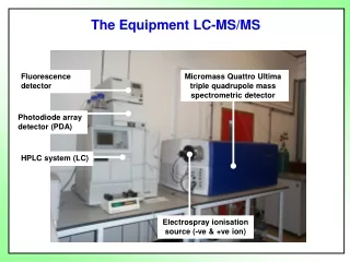
The Equipment LC-MS/MS. Fluorescence detector. Micromass Quattro Ultima triple quadrupole mass spectrometric detector. Photodiode array detector (PDA). HPLC system (LC). Electrospray ionisation source (-ve & +ve ion). A little bit about triple quadrupole mass spectrometry.

E N D
The Equipment LC-MS/MS Fluorescence detector Micromass Quattro Ultima triple quadrupole mass spectrometric detector Photodiode array detector (PDA) HPLC system (LC) Electrospray ionisation source (-ve & +ve ion)
Sample infusion Mass analyser-2 OFF Mass analyser-1 Electrospray source Collision cell Photomultiplier detector Infusion pump n+ Protein (n+x)+ Protein Response Parent ions • Measurement of protein mass by triple quadrupole MS. Mass spectrometer Protein (pure) sample (prepared by investigator) • Multiple-charged ion series - deconvolution gives molecular masses • Intractable analysis for complex protein mixtures. Limited mass resolution.
Applications: Peptide mapping of haemoglobin modified by methylglyoxal
HPLC Mass analyser-2 Mass analyser-1 Electrospray source Collision cell Photomultiplier detector + + Biomarker fragment Enzymatic hydrolysate (prepared by investigator) Response Biomarker Parent ion Fragment ion • Detection of protein biomarkers by LC-MS/MS: • Multiple reaction monitoring (MRM) Mass spectrometer • High specificity • (LC, MS1 and MS2 resolution) • High sensitivity • Biomolecule compatible • Biomarker screening in 75 min per sample.
Advanced glycation endproducts LC-MS/MS with stable isotope-substituted internal standards
Detection of protein biomarkers by LC-MS/MS: Calibration, sample de-lipidification, ultrafiltration & enzymatic hydrolysis • Internal standardisation and calibration • Standards and stable isotope-substituted standards e.g. CML and [13C6]CML, MG-H1 and [15N2]MG-H1 • Delipidification and AGE fractionation • Ultrafiltration to separate protein AGE residues and free AGEs • Ether or methanol/chloroform extraction • Enzymatic digestion: • Pepsin (+ thymol) • Pronase E (under nitrogen, penicillin and streptomycin added) • Prolidase and aminopeptidase (under nitrogen) • Analytical performance • Limits of detection: 20 – 500 fmol. • Recoveries: >80%; 94-100% for amino acids • Interbatch c.v.: <10% (n = 6)
Detection of protein biomarkers by LC-MS/MS: • Retention of amino acids and AGEs and use of column switching To MS/MS Sample Hypercarb column (2.1 x 50 mm) Hypercarb column (2.1 x 250 mm) Switching valve Non-volatile salts to waste • Hypercarb graphitic columns retain underivatised amino acids, allowing for diversion of non-volatile salts to waste. • Column switching facilitates elution of hydrophobic analytes and column washing.
Examples of detection by multiple reaction monitoring (MRM): CML CML detected in plasma protein of a normal healthy human control subject.
Examples of detection multiple reaction monitoring (MRM): Methylglyoxal-derived hydroimidazolone MG-H1 detected in rat retinal protein hydrolysate of a STZ diabetic rat.
Mass spectrometric multiple reaction monitoring detection of protein biomarkers Analyte Rt Parent Ion Fragment ion CE Natural Fragment loss (min) (Da) (Da) (eV) Arg 14.2 175.2 70.3 15 H2CO2, NH2C(=NH)NH2 Lys 6.0 147.1 84.3 15 H2CO2, NH3 Met 9.2 150.0 104.2 11 H2CO2 MetSO 7.5 166.1 102.2 14 CH3SOH CML 7.4 204.9 130.1 12 NH2CH2CO2H MG-H 23.7 229.2 114.3 14NH2CH(CO2H)CH2CH=CH2 Pent 16.5 379.3 250.4 22 NH2CH(CO2H)CH2CH2CH=CH2
HPLC PDA Mass analyser-2 OFF Mass analyser-1 Electrospray source Collision cell Photomultiplier detector + Peptides Resolution of peptide fragments by LC Parent ions Single ion response for each peptide Tryptic digest of protein sample (prepared by investigator) Biolynx match of peptide M+ with theoretical digest. Locate modified peptide M+ ion Peptide map • Peptide mapping to identify sites of protein modification Peptide mapping to identify glycation sites. Mass spectrometer
Glycation of human serum albumin by methylglyoxal Location of glycation sites by LC-MS peptide mapping • Limited proteolysis of MGmin-HSA and HSA control • Reduction of disulphide bonds with dithiothreitol. • S-Alkylation of cysteine thiols by iodoacetamide. • Digestion with trypsin (and independently with Glu-C for corroboration). MS detection of peptide fragments by LC-MS and quantitation of the MS response • Peptides are partially resolved by HPLC with ODS chromatography and detected by positive ion electrospray MS.
MS detection of peptide fragments by LC-MS and quantitation of the MS response • Peptide responses are normalised to the C-terminal peptide (LVAASQAALGL). • Loss of peptides in MGmin-HSA digest was quantified by the mean normalised peptide response for MGmin-HSA, relative to HSA control (mean c.v. = 11%). This is assumed due to glycation • The glycated peptides were also detected as modified dipeptides (resistant to proteolysis in tryptic maps). Modification of arg-410 Peptide(T52) FQNALLVR
Ion chromatograms for dipeptide T52-53 (containing MG-H1-410) Ion chromatograms for peptide T52 (containing R410) LC-MS/MS peptide mapping can also be used to locate glycation, oxidation and nitration markers Location of MG-H1 residues in human serum albumin modified minimally by methylglyoxal • Predicted mass of T52-53 FQNALLVRMG-H1YTK 1406.9; found peptide mass 1406.8 Da.
R114 R428 R186 R410 R218 Glycation of human serum albumin by methylglyoxal Location of glycation sites by LC-MS peptide mapping Modification hotspot: Arg-410 • Drug binding site 2. • Active site of esterase activity.