Download
1 / 65
710 likes | 901 Vues
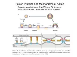
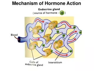
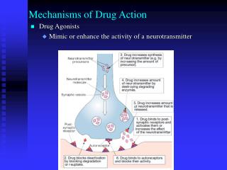
Mechanisms of Hormone Action. General principles:. 1. Signals act over different ranges. 2. Signals have different chemical natures. 3. The same signal can induce a different response in different cells. 4. Cells respond to sets of signals.

E N D
Mechanisms of Hormone Action
General principles: 1. Signals act over different ranges. 2. Signals have different chemical natures. 3. The same signal can induce a different response in different cells. 4. Cells respond to sets of signals. 5. Receptors relay signals via intracellular signaling cascades.
External signals are converted to Internal Responses • Cells sense and respond to the environment Prokaryotes: chemicals Humans: light - rods & cones of the eye sound – hair cells of inner ear chemicals in food – nose & tongue • Cells communicate with each other Direct contact Chemical signals
General Principles of Signal Transduction Signal transduction refers to the overall process of converting extracellular signals into intracellular responses . Key players in signal transduction are signaling molecules, receptors, signal transduction proteins and second messengers, and effector proteins.
Cells respond to signals by changing the activity of existing enzymes (fast) and/or the levels of expression of enzymes and cell components (slower) by gene regulation (Steps 7a & 7b). Receptors and signal transduction systems have evolved to detect and respond to hormones, growth factors, drugs & neurotransmitters.
EXTRACELLULAR FLUID CYTOPLASM Plasma membrane 3 2 1 Reception Transduction Response Receptor Activation of cellular response Relay molecules in a signal transduction pathway Signal molecule Secondary Messengers Target Enzymes Primary Messenger Cascade Effect
- activates an enzyme activity, processes 100 substrates /second primary signal Each protein in a signaling pathway • Amplifies the signal by activating multiple copies of the next component in the pathway Primary enzyme activates 100 target enzymes Each of the 100 enzymes activates an additional 100 downstream target enzymes Each of the 10,000 downstream targets activates 100 control factors so rapidly have 1,000,000 active control factors
Reception Binding of epinephrine to G-protein-linked receptor (1 molecule) Transduction Inactive G protein Active G protein (102 molecules) Inactive adenylyl cyclase Active adenylyl cyclase (102) ATP Cyclic AMP (104) Inactive protein kinase A Active protein kinase A (104) Inactive phosphorylase kinase Active phosphorylase kinase (105) Inactive glycogen phosphorylase Active glycogen phosphorylase (106) Response Glycogen Glucose-1-phosphate(108 molecules) 1 A Signal Cascade amplification 102 104 105 106 108
Receptors relay signals via intracellular SIGNALING CASCADES amplification
Main Types of Receptors ION CHANNEL RECEPTORS G-PROTEIN-COUPLED RECEPTORS KINASE-LINKED RECEPTORS INTRACELLULAR RECEPTORS
Cell-surface receptors -large &/or hydrophilic ligands ion-channel-linked Trimeric G-protein-linked enzyme-linked (tyrosine kinase)
Signal transduction • Control of ion channels • Receptor protein is part of an ion channel protein complex • Receptor binds a messenger leading to an induced fit • Ion channel is opened or closed • Ion channels are specific for specific ions (Na+, Ca2+, Cl-, K+) • Ions flow across cell membrane down concentration gradient • Polarises or depolarises nerve membranes • Activates or deactivates enzyme catalysed reactions within cell
Gate closed Signalmolecule(ligand) Ions Ligand-gated ion channel receptor Plasma Membrane Gate open Cellularresponse Gate close Examples: Muscle Contraction Nerve Cell communication Ion channel receptors
Review: Remember the Na+/K+ ATPase (Na+/K+ pump)? [Na+] inside ~10mM; outside ~150mM [K+] inside ~100mM; outside ~5mM cell has membrane potential ~ -60mV Na+ Cl- -60mV K+ A- - + - - - + - + - + + +
Intercellular Communication When a ligand binds to a receptor protein, the cell has a response. signal transduction: the events within the cell that occur in response to a signal Different cell types can respond differently to the same signal.
Intercellular Communication A cell’s response to a signal often involves activating or inactivating proteins. Phosphorylation is a common way to change the activity of a protein. protein kinase – an enzyme that adds a phosphate to a protein phosphatase – an enzyme that removes a phosphate from a protein
Signal Transduction Components: Kinases/Phosphatases Proteins that participate in intracellular signal transduction fall into two main classes--protein kinases/phosphatases and GTPase switch proteins. Kinases use ATP to phosphorylate amino acid side-chains in target proteins. Kinases typically are specific for tyrosine or serine/threonine sites. Phosphatases hydrolyze phosphates off of these residues. Kinases and phosphatases act together to switch the function of a target protein on or off .
Kinases - Phosphorylation Phosphatase - Dephosphorylation Tyrosine-OH Tyr-Kinases Serine-OH Ser/Thr-Kinases Threonine-OH „dual specificity“ Kinases
There are about 600 kinases and 100phosphatases encoded in the human genome. Activation of many cell-surface receptors leads directly or indirectly to changes in kinase or phosphatase activity. Note that some receptors are themselves kinases (e.g., the insulin receptor).
GH GH binding & dimerisation Binding Activation and of kinases phosphorylation GH receptors (no kinase activity) ATP ADP PO HO OH OP OP OH OP OH kinases HO OH OH OH Growth hormone binding site Kinase active site Growth hormone receptor Tetrameric complex constructed in presence of growth hormone Kinase active site opened by induced fit
Intracellular Receptors steroid hormones -have a nonpolar, lipid-soluble structure -can cross the plasma membrane to a steroid receptor -usually affect regulation of gene expression An inhibitor blocks the receptor from binding to DNA until the hormone is present.
CO2H Steroid binding region Zinc DNA binding region (‘zinc fingers’) H2N Intracellular receptors • Chemical messengers must cross cell membrane • Chemical messengers must be hydrophobic • Example-steroids and • steroid receptors Zinc fingers contain Cys residues (SH) Allow S-Zn interactions
Intracellular Receptors A steroid receptor has 3 functional domains: 1. hormone-binding domain 2. DNA binding domain 3. domain that interacts with coactivators to affect gene expression
Structure of GPCRs G protein-coupled receptors (GPCRs) are the most numerous class of receptors in most eukaryotes. Receptor activation by ligand binding activates an associated trimeric G protein, which in turn interacts with downstream signal transduction proteins. All GPCRs are integral membrane proteins that have a common 7 transmembrane segment structure . The hormone/ligand binding domain is formed by amino acids located on the external side of the membrane and/or membrane interior . GPCRs interact with G proteins via amino acids in the C3 and C4 cytoplasmic regions.
Biological functions mediated by 7TM receptors Smell Taste Vision Neurotransmission Hormone secretion Exocytosis Control of blood pressure Embryogenesis Cell growth and differentiation Development Viral infection Carcinogenesis
G Protein Activation of Effectors The trimeric G protein cycle of activity in hormone-stimulated GPCR regulation of effector proteins is summarized in (next slide).
Trimeric G Proteins & Their Effectors There are 21 different Ga proteins encoded in the human genome. The G proteins containing these subunits are activated by different GPCRs and regulate a variety of different effector proteins . The most common effectors synthesize second messengers such as cAMP, IP3, DAG, and cGMP. In the case of cAMP, a stimulatory Gs subunit activates adenylyl cyclase and cAMP production, whereas an inhibitory Gi subunit inhibits adenylyl cyclase and cAMP production.
GPCRs that Regulate Adenylyl Cyclase Adenylyl cyclase is an effector enzyme that synthesizes cAMP. Ga-GTP subunits bind to the catalytic domains of the cyclase, regulating their activity. Gas-GTPactivates the catalytic domains, whereas Gai-GTPinhibits them. A given cell type can express multiple types of GPCRs that all couple to adenylyl cyclase. The net activity of adenylyl cyclase thus depends on the combined level of G protein signaling via the multiple GPCRs. In liver, GPCRs for epinephrine and glucagon both activate the cyclase. In adipose tissue , epinephrine, glucagon, and ACTH activate the cyclase via Gas-GTP, while PGE1 and adenosine inactivate the cyclase via Gai-GTP.
Activation of Gene Transcription by GPCR Signaling GPCRs regulate gene transcription by cAMP and PKA signaling. cAMP-released PKA catalytic domains enter the nucleus and phosphorylate the CREB (CRE-binding) protein, which binds to CRE (cAMP-response element) sequences upstream of cAMP-regulated genes. Only phosphorylated p-CREB has DNA binding activity. p-CREB interacts with other TFs to help assemble the RNA Pol II transcription machinery at these promoters. In liver, glucagon signaling via this pathway activates transcription of genes needed for gluconeogenesis.
G proteins are important signal transducing molecules in cells. "Malfunction of GPCR [G Protein-Coupled Receptor] signaling pathways are involved in many diseases, such asdiabetes, blindness, allergies, depression, cardiovascular defects, and certain forms of cancer. It is estimated that about 30% of the modern drugs' cellular targets are GPCRs." The human genome encodes roughly 800 G protein-coupled receptors, which detect photons of light, hormones, growth factors, drugs, and other endogenous ligands. Approximately 150 of the GPCRs found in the human genome have still-unknown functions. Whereas G proteins are activated by G protein-coupled receptors, they are inactivated by RGS proteins (for "Regulator of G protein signalling"). Receptors stimulate GTP binding (turning the G protein on). RGS proteins stimulate GTP hydrolysis (creating GDP, thus turning the G protein off).
Down-regulation of GPCR/cAMP/PKA Signaling • A number of events contribute to the termination of signaling by a GPCR. These include: • dissociation of the hormone from the receptor, • hydrolysis of GTP by Ga • hydrolysis of cAMP via cAMPphosphodiesterase, • phosphorylation and “desensitization” of receptors by kinases such as PKA and ß-adrenergic receptor kinase (BARK). • In addition, GPCRs can be removed from the membrane by vesicular uptake.
G-protein activation “molecular switch” inactive • (b) Ligand binds • G-protein associates • (c) GDP-GTP exchange • -Subunit dissociates Active G-Protein-GTP -> allosteric modulator of target effector enzyme active
All G-proteins – similar structure/activation • There are TWO broad subclasses of • trimeric G-protein-activated signal • transduction pathways: • depends on theirtarget effector enzymes • A. adenylyl cyclase • B. phospholipase C
First messenger (signal molecule such as epinephrine) Adenylyl cyclase G protein GTP G-protein-linked receptor ATP cAMP Protein kinase A Cellular responses An activated Ga-protein-GTP • Can trigger the formation of cAMP, which then acts as a second messenger in cellular pathways
G-protein-GTP activation of Effector Enzyme adenylylcyclase produces the 2nd messenger cAMP Activated G-protein
Adenylyl Cyclase & Protein Kinase A Adenylyl cyclase is an integral membrane protein that contains 12 transmembrane segments . It also has 2 cytoplasmic domains that together form the catalytic site for synthesis of cAMP from ATP. One of the primary targets of cAMP is a regulatory kinase called protein kinase A (PKA), or cAMP-dependent protein kinase.
PKA exists in two different states inside cells . In the absence of cAMP, the enzyme forms a inactive tetrameric complex in which 2 PKA catalytic subunits are non-covalently associated with 2 regulatory subunits. When cAMP concentration rises, cAMP binds to the regulatory subunits which undergo a conformational change, releasing the active catalytic subunits.
Phosphorylase kinase inactive + P active Protein Kinase A Phosphorylates downstream target enzymes Breaks down Glycogen Into Glucose
What are targets for Protein Kinase A?? cAMP regulated pathways Function target tissue signal Glycogen breakdown muscle epinephrine Glycogen breakdown liver glucagon Heart rate cardiovascular epinephrine Water reabsorption kidney antidiuretic hormone
How to shut it off? No ligand G-protein -subunit is on a timer Inherent GTPase activity Auto Shut-off
How to shut it off? cAMP-phosphodiesterase rapidly cleaves cAMP (so short lived)
What if you can’t turn off cascade? Vibrio cholera - causes cholera Normal gut: H20, NaCl, NaHCO3 secretion controlled by hormones via Gs/cAMP signal pathways V. cholera – secretes enterotoxin, chemically modifies Gs – no GTPase activity - stays ON Severe watery diarrhea – dehydration, death
target effector enzyme is Phospholipase C PLC cleaves a membrane phospholipid (Phoshatidyl inositol) to two 2nd Messengers: Inositol-1,4,5-Trisphosphate (InsP3) & Diacylglycerol (DAG)