Download
1 / 69
961 likes | 2.02k Vues
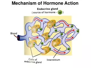
Hormone Biosynthesis, Metabolism, and Mechanism of Action. Adrian Quesada Rojas, MD. Definitions. Hormone: Substance produced in tissue => bloodstream => responsive cells Provides means of communication chemical regulatory and signaling Bloodstream Paracrine: cell to cell (contiguos)

E N D
Hormone Biosynthesis, Metabolism, and Mechanism of Action Adrian Quesada Rojas, MD
Definitions Hormone: Substance produced in tissue => bloodstream => responsive cells Provides means of communication chemical regulatory and signaling Bloodstream Paracrine: cell to cell (contiguos) Autocrine: same cell Intracrine: same cell (unsecreted)
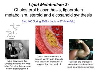
Nomenclature Steroid hormones Sex steroids (Cholesterol derivatives) 3 groups (number of carbon atoms) 21 carbons: pregnane nucleus 19 carbons: androstane 18 carbons: estrane
Lipoproteins and Cholesterol • All steroid prod organs can synth (acetate) LPP: transport of non polar fat in polar solvent • Chylomicrons: chlol 10, TG 90 formed in GI • VLDL: more dense • IDL: removal of some TG from VLDL interior • LDL: end prod VLDL catabolism (50% chol) • major carrier of chol in plasma • HDL: highest protein content
Steroidogenesis • Reactions: Cleavage of side chain Conv hydroxyl <=> ketones (dehydrogenase) Addition of OH group (hydroxylation) Creation of double bonds (remov of H) Addition of hydrogen (saturation) Enz are dehydrogenases or C P450 oxidases
Steroidogenesis • Rate limit step: transfer Chol from outer to inner mitochondrial membrane (SSC) • Once Pregnenolone: two ways in ovary D5pathway (3-b hydroxysteroids) => DHEA D4pathway (3-ketone) => 17a hydroxyP • Conversion of pregnenolone to P: 3b hydroxysteroid dehydrogenase D4-5isomerase reaction (3-OH group to ketone + transfer double bond 5-6 to 4-5)
Steroidogenesis • P hydroxylated at 17 position 17a OH P (precursor of C19 androgens) By peroxide formation at C20, followed by epoxidation of C17, C20 carbons, side chain is split off forming Androstenedione • 17 ketone may be reduced to 17b OH to form testosterone (C 19) • C 19 steroids => aromatase => C18 phenolic steroid estrogen (E1 and E2)
Steroidogenesis • Alternative Pregnenolone conv to D5 3b OH C19 steroid (DHA) by 17a OH lation followed by SCC With D4 3 ketone formation, DHA is converted into Androstenedione (C19 steroids) C 19 steroids undergo aromatization (hydroxylation of angular 19 methyl group, followed by oxydation and loss of 19 C as formaldehyde and dehydrogenation)
Two Cell System • FSH receptors on granulosa • FSH receptors are induced by FSH • LH receptors on theca initially, as follicle grows FSH induces LH receptors on granulosa • FSH ind aromatase activity on granulosa • Actions regulated by autocrine and paracrine factors
Blood Transport of Steroids • Most E and T bound to protein carrier (SHBG) • 30% is bound to albumin • 1% free Hyperthyroidism, pregnancy, E adm SHBG Corticoids, A, P, GH, Insulin SHBG SHBG inversely related to weight Hyperinsulinemia (low SHBG predictor of DM) Body fat distrib (central=> hyperinsulinemia)
Estrogen Metabolism • Ovary => Estradiol / estrone • Estriol => periph metab of estrone / E not a secretory prod of ovary conversion to less active form Androgens are precursors of estrogens Adrenal gland (source of A, Androstenedione)
Estrogen Metabolism • E2 100-300 mg/day • Androstenedione 3 mg/day ( 1% conversion) => 20- 30 % of E1 (produced every day)
Progesterone Metabolism • NO peripheral conversion • Secretion from adrenal and ovaries • Preov < 1 mg /day • Post ov 20-30 mg /day • 10-20% of P is excreted as Pregnanediol • Pre ov <1 mg/day, post ov 3-6 mg/d (home ovulation test)
Androgen Metabolism • Ovary: DHA, Androstenedione, testosterone • Adrenal cortex: Gluco, mineralo, sex steroid • Sex steroids (less than ovary) • DHA (½ adrenal, ¼ ovary, ¼ periph) • Androstenedione (½ adrenal, ½ ovary) • Testost 0.2-0.3 mg/day • 50% periph conv of androstenedione and DHA • 25% adrenal • 25% ovary
Androgen Metabolism • Androgens Test binding capacity • Androg effects depends on unbound fraction • Hirsutism • Test => 5a reductase => DHT (princ androg) • Androstenedione > Test (women) • DHT is derived from Androst and DHA • DHT largely metabolized intracell • Only 1/10 of levels of Test • Not all tissue requires DHT from Test (Wolffian)
Cellular Mechanism of Action • Two major types of hormone action Tropic hormones (recep at cell memb) Steroids (recep inside the cell) • Multiple cell receptors: Intracellular: In the nucleus (transcription activity) G protein: Single polypeptide chain (cell memb)
Cellular Mechanism of Action Ion gate channel: Cell surface, mult units (ACh) Intrinsic enz activ: Trans memb recep with intracell component with tyrosine or serine kinase activity
Mechanism of actionSteroid hormones • Includes: • Hormone diffusion across cell memb • Steroid binding to receptor protein • Interaction of Horm-Recep complex with DNA • Synthesis of mRNA • Transport of mRNA to ribosomes • Protein synthesis in cytoplasm
Mechanism of actionSteroid hormones • Biological activity Mantained only while H-R complex attached to nuclear site Duration of exposure to hormone is as important as dose Major factor in potency differences among various estrogens is length of time E-R complex occupies nucleus
Mechanism of actionSteroid hormones Cortisol and P must circulate in large concentrations 2 to receptor complexes short half-lives in nucleus H-R complex after gene activation (processing) Rapid degradation of R unbound to E Much slower degradation of bound R Continuous presence of E is important factor in continuing response
Estrogen Receptors • ER-a (rapid turnover) • ER-b 96% homologous in DNA binding domain and 53% homologous in the hormone binding domain (when compared to ER-a) • Respond in comparable manner to same hormones
Estrogen Receptors • E receptors => nucleocytoplasmic shuttling • Recep can diffuse out of nucleus and be transported back in or undergo metabolism • If shuttling impaired Receptors are degraded rapidly • E antagonists prevent nuclear translocation and thus increase cytoplasmic degradation
Estrogen Receptors • Prior to binding R is inactive complex that includes a variety of heat shock proteins • Activation => dissociation of HSP • When H binds R => conformational change • Conformational shape determines exact message transmitted to the gene • E2, raloxifene, tamoxifen induces a different conformational shape
Estrogen Receptors • Once activated H receptor activates transcription in partnership with several groups of polypeptides • Transcription factors (polymerase enzyme and DNA) • Coactivators and corepressors (intracel prot called adaptor proteins and activate or suppress the TAF)
Estrogen Receptors • Phosphorylation • Also stimulated by ligand binding • Occur in specific receptor sites • Increases potency of molecule to regulate transcription • cAMP and prot kinase pathways increase transcriptional activity of E-R
Estrogen Receptors • Differences ER-a and ER-b ER-b prevalent in brain, CV system, granulosa cells Breast expresses ER-a and ER-b ER-b acts as natural suppressor of ER-a activity on breast tissue Colon contains only ER-b, reduction of colon CA in postmenopausal on HRT may reflect antiproliferative effect of beta receptor
Progesterone Receptor • Induced by E, decreased by Progestins • Two major forms (A and B) • A and B are expressed differently in Breast and Endometrial tissues • In most cells B positive regulator of P-responsive genes, A inhibits B activity • A and B have different molecular functions, affecting different genes
Progesterone Receptor • Mice lacking of P receptors (A,B) are unable to ovulate (failure to expel a mature oocyte in a fully developed follicle) • PR-A protects against uterine and mammary gland hyperplasia induced by PR-B
Androgen Receptor • Androgens only in one of three ways: • T => DHT (Intracrine) • T itself (Endocrine) • Intracel conversion of T to E (Intracrine) • Wolffian duct derivatives (T) • Hair follicles, urogenital sinus derivatives (req T => DHT)
Androgen Receptor • T and DHT binds to same high affinity A-receptor (DHT has greater affinity) • Anti-A also bind to same R (20% affinity of T) • A-R: two forms B (full length) and A (shorter) • Functional differences yet to be determined • A and P can cross react for their receptors (pharmacologic concentrations) • Progestin can act as an anti-E and anti-A
Androgen Receptor • Androgen Insensitivity Syndrome Congenital abnormality in A intracel R A-R gene is on X chromosome (Xq11-12) X-linked disorder Deletion of amino acids from steroid binding domain 2 to nucleotide alterations in gene
Agonists and Antagonists Agonist => stimulates response Antagonist => inhibits actions of an agonist blockage of receptor message • Examples: Tamoxifen Mifepristone Histamine receptor antag
Agonists and Antagonists • Physiologic Antagonists P is not an E antagonist Modifies E action by depleting E receptors P can induce conversion of E2 to E1 Androgens block E actions (unclear mechan)
Agonists and Antagonists • Two groups of anti-E: Pure anti-E Mixed agonists antagonists • Tamoxifen: Similar to clomiphene Competitively inhibits E binding to receptor E affinity for receptor is 100-1000x better than tamoxifen
Agonists and Antagonists • Tamoxifen–E receptor binds to DNA • Agonistic – Antagonistic actions depends on coactivators present in specific cell types • Estrogenic actions: • Lowers FSH levels • Decreases Cholesterol, LDL • Stimulates P receptor synthesis • Maintenance of bone, vagina, endometrium
Agonists and Antagonists • Causes endometrial hyperplasia, polyps and 4x increase in endometrial cancer • Mechanism of action: • TAF-1 and TAF-2 can both activate transcription • Tamoxifen agonistic ability is due to activation of TAF-1 • Antagonistic activity is due to competitive inhibition of E-dependent activation of TAF-2
Agonists and Antagonists • Response to E and anti-E depends on: • Nature of E receptor • E response elements and promoters • Cell content of protein coactivators • Properties of ligand • Modulation by growth factors and agents that affect phosphorylation and protein kinases
Agonists and Antagonists • Tamoxifen reduces risk of recurrent disease in E-R positive and negative breast cancer • Besides binding to E-R (competitive inh), • Inhibits protein kinase C activity (phosphor) • Inhibits calmodulin-dependent cyclic AMP phosphodiesterase (binds to calmodulin) • Stim secretion of TGF-b (inh growth CA cell)
Agonists and Antagonists • Tamoxifen: treatment should be for only 5 years • Emergence of tamoxifen-resistant tumors, how? Loss of E receptors • Loss of cellular control / loss of E-R expression • Variant and mutant E-R • Changes in coactivators • Differential metabolism • Resistance occur when E-R is NOT dominant in growth of CA cells