Download
1 / 23
230 likes | 336 Vues
Explore the latest developments in imaging and understanding magnetic nanostructures using X-rays with ultrafast time scales and nanoscale resolution. Discover key insights into magnetization control and dynamics at the atomic level.

E N D
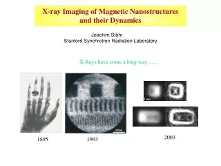
X-ray Imaging of Magnetic Nanostructures and their Dynamics Joachim Stöhr Stanford Synchrotron Radiation Laboratory X-Rays have come a long way…… 1 mm 2003 1895 1993
Andreas Bauer1,2 Jan Lüning2 Andreas Scholl1 Howard A. Padmore1 Yves Acremann2 Aaron Lindenberg3 Andrew Doran1 Sug-Bong Choe1 Hendrik Ohldag2 Squaw Valley, April 2003 1 Advanced Light Source 2 Stanford Synchrotron Radiation Laboratory 3 UC Berkeley
Fe metal – L edge Kortright and Kim, Phys. Rev. B 62, 12216 (2000) Soft X-Rays are best for magnetism!
Transmission X-ray Microscope Reconstruction from Speckle Intensities Imaging by Coherent X-Ray Scattering Phase problem can be solved by “oversampling” speckle image 5 m (different areas) S. Eisebitt, M. Lörgen, J. Lüning, J. Stöhr, W. Eberhardt,E. Fullerton (unpublished)
s s [010] 2m m Spectromicroscopy of Ferromagnets and Antiferromagnets AFM domain structure at surface of NiO substrate FM domain structure inthin Co film on NiO substrate NiO XMLD Co XMCD H. Ohldag, A. Scholl et al., Phys. Rev. Lett. 86(13), 2878 (2001).
Magnetic characterization of interfacial spins Co/NiO Co/IrMn Co NiO Publications: Stöhr et al., Phys. Rev. Lett. 83, 1862 (1999) Thomas et al., Phys. Rev. Lett. 84, 3462 (2000) Scholl et al., Science 287, 1014 (2000) Nolting et al., Nature 405, 707 (2000) Regan et al. Phys. Rev. B 64, 214422 (2001) Ohldag et al., Phys. Rev. Lett. 86 2878 (2001) Ohldag et al., Phys. Rev. Lett. 87, 247201 (2001) Ohldag et al., Phys. Rev. Lett. 91, 017203 (2003) loop of interfacial spins - only 4% are pinned
ideal AFM poly AFM Exchange Bias Model from X-Rays
Present limitations of magnetic recording • Present method of magnetic switching is unfavorable: • present recording time ~1 ns • unfavorable torque and dependent on thermal activation
Fast Magnetization Dynamics is governed by Landau-Lifschitz-Gilbert equation: Angular momentum change Precession torque Gilbert damping torque 1 Tesla field: 90o rotation in 10 ps Typically a << 1, 100 ps We want to understand a on atomic level a controls switching time, a~1 optimal
Time Resolved X-Ray Microscopy Laser pump – x-ray probe synchronization < 1 ps excitation laser pulse < 100 ps observation x-ray pulse t 328 ns
100 m 100 m 10 m 2 m 2 m Production of Magnetic Field Pulses Photoconductive switch H ~ 200 Oe Conducting wire 50 => I = 200 mA, 10 V bias Magnetic Cells Current
Sample and Magnetic Field Pulse 20 nm Co90Fe10 films with in-plane anisotropy (1 m) x (1-3 m) rectangles Current magnetic field Magnetic Field Pulse ~ 150 Oe at Maximum < 50 ps rising time > 300 ps decaying time with some reflection M
Observation of Vortex Motion H 1 mm x 1 mm 2 mm x 1 mm 1.5 mm x 1 mm • Vortices rotate oppositely • vortex cores point in opposite directions Vortex speed ~ 100 m/s
Conclusions The challenge of the future is to control the magnetization on the nanometer length scale and picosecond/femtosecond time scale Our current capabilities are: • image the magnetization with 50 nm spatial resolution, • image the response of the magnetization with 100 ps time- and 100 nm spatial resolution Outlook into the future: • 5 nm spatial resolution – PEEM3, under construction • 100 fs time resolution: pump-probe excitations single snapshots of equilibrium dynamics Modern x-ray sources offer unique opportunities for studies of the ultrafast magnetic nanoworld
Vortex Structure And Vortex Motion Elevation view torque H Plane view Motion antiparallel to field! Landau-Lifshitz equation: (neglect damping) The field acts like a screw driver. Depending on the orientation of the thread pitch, the screw (vortex) will move either forward or backward
x dx dH H Vortex Precession Under a field pulse, the vortex moves from the center. M Happlied After the field pulse, the vortex continues to move radially due to the magnetostatic energy. Induced magnetostatic field is always perpendicular to the vortex motion. Hmagnetostatic Magnetostatic field is always perpendicular to the vortex deviation Vortex will precess forever if there is no damping.
scattering vector q (mm-1) scattering vector q (mm-1) 40 40 20 20 0 0 scattering vector q (mm-1) -20 -20 -40 -40 -40 -40 -20 -20 0 0 20 20 40 40 scattering vector q (mm-1) Incoherent vs. Coherent X-Ray Scattering Small Angle Scattering Coherence length larger than domains, but smaller than illuminated area information about domain statistics Speckle Coherence length larger than illuminated area true information about domain structure
Pulse Structure Possible solutions: - gated detector, pulse picker - pump at 500 MHz