LEISHMANIASIS
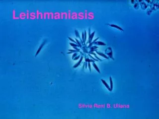
LEISHMANIASIS. Leishmaniae are protozoans transmitted to humans by insect bites and cause a spectrum of clinical syndromes, ranging from indolent ,self resolving cutaneous ulcers to fatal disseminated disease


LEISHMANIASIS
E N D
Presentation Transcript
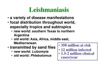
Leishmaniae are protozoans transmitted to humans by insect bites and cause a spectrum of clinical syndromes, ranging from indolent ,self resolving cutaneous ulcers to fatal disseminated disease • Trnsmitted by phlebotomus sandflies,which acquire the infection by feeding on infected animals.
It is mainly a disease of less developed countries where humans live in close proximity to animal hosts. • sandfly • inoculation of organisms into the skin .
phagocytosis by macrophages • transform into Amastigotes
reproduce within the macrophages • rupture of the cell,infect other macrophages • cluster of infected macrophages at the site of inoculation
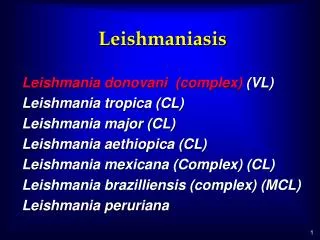
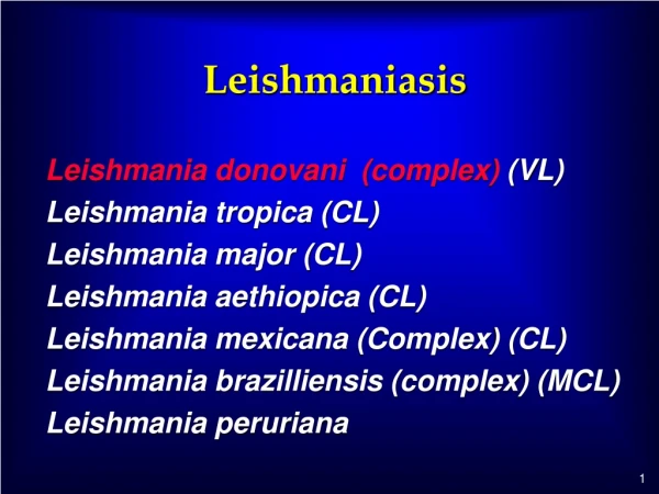
Localized Cutaneous Leishmaniasis • Disseminated Cutaneous Leishmaniasis • Mucocutaneous Leishmaniasis • Visceral Leishmaniasis.
Localized Cutaneous Leishmaniasis: Leishmania species in Central &South America,Northern Africa,the Middle East,India,and china produce localized cutaneous disease,also known as oriental sore or tropical sore.
Localized CutaneousLeishmaniasis Begins as a collection of amastigotes-filled macrophages that ulcerates the overling epidermis. In tissue sections, the oval amastigotes measure 2micrometer and contain two internal structures – a nucleus and a kinetoplast. L.D bodies ---Under low power amastigotes in macrophages appear as dots.
With progreesive develpoment of cell- mediated immunity macrophages become activatedand kill the bacteria. • Slowly a granulomatous appearance • Over the course of months,the cutaneous ulcers heals spontaneously.
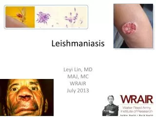
Clinical features: • Cutaneous Leishmaniasis:begins as itching, solitary papule ,which erodes to form a shallow ulcer with sharp raised border with crust formation. • These lesions grow up to 3-6cm.
Diffuse CutaneousLeishmaniasis • Develops in some patients who lack specific cell-mediated immune responses to leishmaniae. • The disease begins as a single nodule, but adjacent satelite nodules slowly form,evantually involving much of the skin. • Closely resemble lepromatous leprae.
Caused by L.Braziliences • Early course and pathologic changes are similar to those of localized cutaneous L. • A solitary ulcer develops resolves spontaneously. • Years after primary lesion has healed,an ulcer develops at a mucocutaneous junction;larynx,nasal septum,anus,vulvaetc
Destruction of nose--------tapir nose • Obstruction of the airways.
VISCERAL LEISHMANIASIS(KALA AZAR) • Produced by several species of Leishmania Donovani • Also called Black fever • Involves reticuloendothelial system