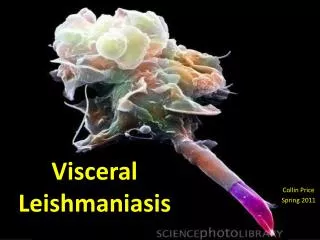
Leishmaniasis
Leishmaniasis . Dr.T.V.Rao MD. Sir William Leishman . 1900 – Sir William Leishman discovered L. donovani in spleen smears of a soldier who died of fever at Dum-Dum, India. The disease was known locally as Dum-Dum fever or kala-azar. Charles Donovan .

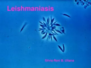
Leishmaniasis
E N D
Presentation Transcript
Leishmaniasis Dr.T.V.Rao MD Dr.T.V.Rao MD
Sir William Leishman • 1900 – Sir William Leishman discovered L. donovani in spleen smears of a soldier who died of fever at Dum-Dum, India. The disease was known locally as Dum-Dum fever or kala-azar. Dr.T.V.Rao MD
Charles Donovan • Charles Donovan also recognized these symptoms in other kala-azar patients and published his discovery a few weeks after Leishman. After examining the parasite using Leishman's stain, these amastigotes were known as Leishman-Donovan bodies. Dr.T.V.Rao MD
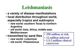
Leishmaniasis is Neglected Disease • Leishmaniasis is a globally important but neglected disease, affecting approximately two million people every year. For most people, infection results in a slow-to-heal skin ulcer. In others, however, the parasite targets the liver, spleen and bone marrow, leading to over 70,000 deaths annually. Dr.T.V.Rao MD
Phylum Order Family Genus Sarcomastigophora Kinetoplastida Trypanosomatidae Leishmania The Parasite Dr.T.V.Rao MD
Leishmania Parasites and Diseases Dr.T.V.Rao MD
Promastigote Amastigote Morphology Flagella Kinetoplast Golgi Nucleus Cytoskeleton Dr.T.V.Rao MD
Morphology and Life Cycle • Amastigotes measure 2-3 micrometers, with a large nucleus and Kinetoplast. • Amastigotes mainly live within cells of the RE system, but have been found in nearly every tissue and fluid of the body. Dr.T.V.Rao MD
Life cycle • The organism is transmitted by the bite of several species of blood-feeding sand flies (Phlebotomus) which carries the Promastigote in the anterior gut and pharynx. It gains access to mononuclear phagocytes where it transform into Amastigote and divides until the infected cell ruptures. Dr.T.V.Rao MD
Sand fly- Vector Dr.T.V.Rao MD
Life cycle • The released organisms infect other cells. The sand-fly acquires the organisms during the blood meal, the Amastigote transform into flagellate Promastigote and multiply in the gut until the anterior gut and pharynx are packed. Dogs and rodents are common reservoirs. Dr.T.V.Rao MD
Different stages of Haemoflagellates Dr.T.V.Rao MD
1- Sand-fly bites animal and ingests blood infected with Leishmania 2- Sandfly bites human and injects Leishmania into skin 4- Cycle continues when sandfly bites another human or animal reservoir 3- Another sandfly bites human and ingests blood infected with Leishmania
What is Kala-Azar • Kala-azar means dark pigmentation which is characteristic of cases of visceral leishmaniasis. It is caused by Leishmania donovani bodies and may be present either in endemic, epidemic or sporadic forms. It is widely prevalent in India in epidemic form in states of Bihar, Assam and Bengal. Kala azar found in East and North Africa is a disease of young children and young adults, being more common in males as compared to females. Dr.T.V.Rao MD
Clinical types of cutaneous leishmaniasis • Leishmania major:Zoonotic cutaneous leishmaniasis: wet lesions with severe reaction • Leishmania tropica:Anthropologic cutaneous leishmaniasis: Dry lesions with minimal ulceration Oriental sore(most common) classical self-limited ulcer Dr.T.V.Rao MD
KALA AZAR • Leishmaniasis is a disease caused by protozoan parasites of to the genus Leishmania and is transmitted by the bite of sand fly. • This disease is also known as kala azar, black fever, sandfly disease, Dum-Dum fever. • Human infection is caused by about 21 of 30 species that infect mammals. These include the L. donovani complex with three species (L. donovani, L. infantum, and L. chagasi
Cutaneous leishmaniasis Dr.T.V.Rao MD
Diffuse cutaneous leishmaniasis Leishmaniasis recidiva Dr.T.V.Rao MD
Uncommon types • Diffuse cutaneous leishmaniasis (DCL): Caused by L. aethiopica, diffuse nodular non-ulcerating lesions. Low immunity to Leishmania antigens, numerous parasites. • Leishmaniasis recidiva(lupoid leishmaniasis): Severe immunological reaction to Leishmaniaantigen leading to persistent dry skin lesions, few parasites. Dr.T.V.Rao MD
Post Kala-azar Dermal Leishmaniasis • Post Kala-azar Dermal Leishmaniasis (PKDL) is a condition when Leishmania donovani invades skin cells, resides and develops there and manifests as dermal leisions. Some of the kala-azar cases manifests PKDL after a few years of treatment. Recently it is believed that PKDL may appear without passing through visceral stage. Dr.T.V.Rao MD
Pathogenesis • Infections range from asymptomatic to progressive, fully developed kala-azar. • Incubation period is usually 2 – 4 months. • Symptoms – Begins with low-grade fever and malaise, followed by progressive wasting, anemia, and protrusion of the abdomen from enlarged liver and spleen. • Fatal after 2 – 3 years if not treated. • In acute cases with chills, fevers up to 104 degrees Fahrenheit, and vomiting; death may occur within 6 – 12 months. • Immediate cause of death is usually an invasion of a secondary pathogen that the body is unable to combat. Dr.T.V.Rao MD
Cutaneous leishmaniasis Diagnosis: • Smear: Giemsa stain – microscopy for LD bodies (Amastigote) • Biopsy: microscopy for LD bodies or culture in NNN medium for promastigotes Dr.T.V.Rao MD
L. donovani bodies • L. donovani bodies may be demonstrated in buffy coat preparations of blood and bone marrow aspirate. Aspirates taken from enlarged lymph nodes show parasites in 60 percent of cases. Dr.T.V.Rao MD
Visceral leishmaniasis Diagnosis • Parasitological diagnosis: METHOD Bone marrow aspirate 1. microscopy Splenic aspirate 2. culture in NNNmedium Lymph node Tissue biopsy Dr.T.V.Rao MD
Bone marrow aspiration Bone marrow amastigotes Dr.T.V.Rao MD
Diagnostic Methods in Leishmaniasis • Antibody detection. Specific sero diagnostic tests are also employed. Conventional methods include gel diffusion, complement fixation test, indirect haem agglutination test, indirect immuno-fluorescent antibody test (IFAT) and counter immuno electro phoresis. Most of these tests have limited sensitivities and specifies. Dr.T.V.Rao MD
Culturing of the Parasite • Organisms can be cultured in Nicolle-NovyMacneal (NNN media) media from clinical specimens obtained from splenic or bone marrow aspirates. Dr.T.V.Rao MD
Immunological Diagnosis: • Specific serologic tests: Direct Agglutination Test (DAT), ELISA, IFAT • Skin test (leishmanin test) for survey of populations and follow-up after treatment. • Non specific detection of hypergammaglobulinaem by formaldehyde (formal-gel) test or by electrophoresis. Dr.T.V.Rao MD
Direct agglutination test • Direct agglutination test (DAT) based on agglutination of the trypsenized whole promastigotes is useful in endemic regions. Its sensitivity ranges from 91-100% and specificity from 72 to 100%. Dr.T.V.Rao MD
ELISA • ELISA is an important sero diagnostic tool for leishmaniasis. It is a highly sensitive test and its specificity depends upon the antigen used. Dr.T.V.Rao MD
Chromatographic strip test • A ready to use immuno chromatographic strip test based on rk 39 antigen has been developed as a rapid test for diagnosis of kala azar. An important limitation of this test is the presence of antibodies in healthy controls hailing from endemic regions. Dr.T.V.Rao MD
Treatment: • Pentavalent antimony (Pentostam) • Amphotericin B Treatment of complications: • Anemia • Bleeding • Infections etc. Dr.T.V.Rao MD
Management of Kala-azar Patients • It includes both supportive and curative. All patients of Kala azar should preferably be hospitalized. Any infection complicating the disease be treated by use of proper antibiotics. Nutrition must be maintained. Cases with severe anemia may require blood transfusions. Pentavalent antimony compounds are the drug of choice. Sodium antimony gluconate (Pentostam) is the most commonly used drug. Dr.T.V.Rao MD
Kala-azar prevention: • Multipronged approach is needed. • Sand-flies are extremely sensitive to insecticides & vector control through insecticide spray is very important. • Mosquito nets or curtains treated with insecticides will keep out the tiny sand-flies. Dr.T.V.Rao MD
Kala-azar prevention: • In endemic areas with zoonotic transmission, infected or stray dogs should be destroyed. • In areas with anthroponotic transmission, early diagnosis & treatment of human infections, to reduce the reservoir & control epidemics of VL, is extremely important. • Serology is useful for screening of suspected cases in the field. • No vaccine is currently available . Dr.T.V.Rao MD
Programme Created by Dr.T.V.Rao MD for Medical and Paramedical Students in the Developing World • Email • doctortvrao@gmail.com Dr.T.V.Rao MD