Download
1 / 15
150 likes | 200 Vues
Delve into the world of the heart and its functions through interesting notes on cardiovascular anatomy, ECG invention, heart facts, and disorders like heart disease and fibrillation. Learn about treatments like nitroglycerin and pacemaker implantation.

E N D
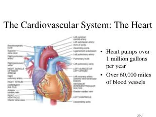
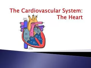
Cardiovascular System Notes: Heart Disease & Disorders
Interesting Heart Facts The Electrocardiograph (ECG) was invented in 1902 by Willem Einthoven Dutch Physiologist.This test is still used to evaluate the heart's rate and rhythm.
Review What are the 3 parts of the cardiovascular system? heart – blood – blood vessels What do arteries do? Take blood AWAY from the heart What are the 3 layers of an artery? Tunica externa /connective tissue (elastin) – tunica media/smooth muscle – tunica intima/endothelium What do veins do? Bring blood TOWARD the heart What do veins have that arteries don’t? VALVES – (veins & arteries both have the same layers except the muscular layer is smaller)
What exchanges material between blood and the body’s cells? CAPILLARIES How big are they? microscopic in size What is the outer membrane of the heart called? Pericardium What is the function of the pericardium? (there are 3) • protection • anchors heart to other structures • provides lubrication for heartbeat
What are the 3 layers of the heart wall? • Epicardium – outside layer • Myocardium – middle layer (cardiac muscle) • Endocardium – inner layer What does the septum do? Divides the heart into right and left sides What are the 4 chambers of the heart? • Right Atria • Left Atria • Right Ventricle • Left Ventricle
What is the function of the right atria? receives blood from inferior & superior vena cava (oxygen poor) What is the function of the left atria? receives blood from pulmonary veins (oxygen rich blood from the lungs) What does the function of the left ventricle ? receives blood from left atria & pumps it to the body (through the aorta) What is the function of the right ventricle? receives blood from the right atria and pumps it to the lungs (through the pulmonary arteries)
Where is the tricuspid valve located? between right atria and right ventricle Where is the bicuspid (mitral) valve located? between left atria and left ventricle Where is the pulmonary semilunar valve located? between pulmonary artery and right ventricle Where is the aortic semilunar valve located? between aorta and left ventricle What is the pacemaker of the heart? Sinoatrial node or SA node (begins each heartbeat)
What does the atrioventricular node (AV node) do? Receives the impulse from the SA node (sinoatrial node) Trace the impulse of a heartbeat beginning at the SA node SA node – AV node – Purkinje Fibers (network) – up the sides of the ventricles What is systole? What is diastole? Systole = contraction of the ventricle Diastole = relaxation of the ventricle What is an electrocardiogram? Amplification of heart’s electric current that produces distinct wave patterns: P wave = depolarization of atria QRS complex = depolarization of ventricle T wave = repolarization of ventricles
HEART DISORDERS & DISEASES • Risk Factors: • cholesterol – diet, genetics • hypertension (high blood pressure) • smoking – excessive drinking • obesity – lack of exercise • heredity
Heart Disease • term covers many different types of heart problems • Coronary Artery Disease (CAD) or Coronary Heart Disease (CHD) • involves inadequate blood supply to heart muscle • can lead to: • ANGINA – severe chest pain due to brief lack of oxygen to heart muscle (can be mistaken for a heart attack)
ISCHEMIA – a decrease in blood supply to heart • INFARCTIONS – dying of tissue due to lack of blood supply and oxygen
Myocardial Infarction • M.I. aka: heart attack • blood supply to the heart is cut off (ischemia) resulting in heart tissue dying dead or dying heart tissue • medical emergency – nitroglycerin (vasodilator) – angioplasty – bypass surgery • Treatments:
Fibrillation • problem with the SA node or AV node • Atrial Fibrillation • atria “quiver” or do not contract normally
Ventricular Fibrillation • very dangerous, death is usually a result
Bradycardia • a slowing of the heart rate due to SA or AV node problems • Treatment: pacemaker implantation to regulate heart beat • Heart Murmur • valves don’t close correctly and blood leaks back through the valve • Treatment: medication, new valve • valves become calcified, rough, narrow and do not close properly • Stenosis