
Anatomy for Complete and Partial Dentures
E N D
Presentation Transcript
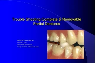
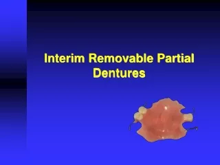
1. Anatomy for Complete and Partial Dentures
2. Lips Vermilion Border
Denture provides lip support
Affects vermilion border width
3. Lips Philtrum
Depression below nose
4. Lips Nasolabial Angle
Angle between columella of nose & philtrum of lip
Normally, approximately 90� as viewed in profile
5. Lips Tissue of the Upper Lip
Loose tissue of the upper lip can be gathered between your thumb and index finger
6. Cheeks Masseter Muscle
Closing muscle bulges into distal corner of buccal vestibule
Not active during impression making
7. Residual Ridges If ridges are severely resorbed, inform patient
�U�-shape
�V�-shape
8. Vestibules If vestibules are shallow, inform the patient
9. Maxilla Maxillary Tuberosities
Oversized
Resorbed
Undercut
10. Maxilla Maxillary Tuberosities
Oversized
Resorbed
Undercut
11. Maxilla Incisive Papilla
Landmark for setting of teeth
12. Maxilla �Hamular� Notch
Posterior border denture
Between the bony tuberosity and hamulus
�Soft displaceable tissue�, for comfort and retention
13. Maxilla �Hamular� Notch
Posterior border denture
Sometimes posterior to where the depression in the soft tissue appears
Use the head of your mirror to palpate the notch & mark with an indelible marker
14. Maxilla Soft Palate
Vibrating Line
Critical posterior border dentures
Junction of movable and immovable portions of the soft palate
15. Maxilla Glandular Tissue
Soft displaceable
16. Maxilla Soft Palate
Fovea Palatine
Bilateral indentations near midline of the soft palate
Close to the vibrating line
17. Maxilla Hard Palate
Median Palatine Raphe (midline palatine suture)
A bony midline structure
May require relief when covered by a denture
18. Maxilla Torus Palatinus
May require removal
19. Mandible Pear Shaped Pad
Soft pad containing glandular tissue
Inverted pear shape, posterior border
Created from scarring after extractions
20. Mandible Buccal Shelf
Primary denture bearing area of mandibular denture
Between height of bridge & external oblique ridge
Resorbs more slowly
21. Mandible Anterior Border of the Ramus
Do not extend dentures to ramus
Discomfort will result
22. Mandible External Oblique Ridge
Do not extend dentures to this ridge
23. Mandible Mylohyoid Ridge
Origin of mylohyoid muscle which influences length of lingual flange
Can be prominent, and/or sharp, requiring relief
24. Mandible Mylohyoid Ridge
25. Mandible Lingual Tori
Raised bony structures
May require relief when covered by a denture
Thin mucosa can ulcerate easily
26. Mandible Genial Tubercles
Attachment for the genioglossus muscle
Tubercles may be higher than the ridge with severe resorption
27. Frena (singular = frenum) Must be relieved to allow movement, without impingement
If prominent, adequate relief can weaken a denture
If too much relief, retention is lost
Check prominence intraorally
28. Pterygo-Mandibular Raphe Connects from the hamulus to the mylohyoid ridge
When prominent, can cause pain, or loosening
Requires relief �groove � if prominent
29. Retrozygomal Fossae (Space) Palpate zygomatic process in buccal vestibule just buccal to first maxillary molar
Vestibular space posterior to zygoma
30. Retrozygomal Fossae (Space) Commonly incompletely captured in preliminary impressions
Use syringe technique
31. Coronoid Process Place mirror head lateral to tuberosity
Move mandible to opposite side
Note binding or pain
This gives some indication of the width of the space for flange