Kidney regeneration
Manifestation of Novel Social Challenges of the European Union in the Teaching Material of Medical Biotechnology Master’s P rogrammes at the University of Pécs and at the University of Debrecen Identification number : TÁMOP-4.1.2-08/1/A-2009-0011.

Kidney regeneration
E N D
Presentation Transcript
Manifestation of Novel Social Challenges of the European Unionin the Teaching Material ofMedical Biotechnology Master’s Programmesat theUniversity of Pécs and at the University of Debrecen Identificationnumber: TÁMOP-4.1.2-08/1/A-2009-0011
Manifestation of Novel Social Challenges of the European Unionin the Teaching Material ofMedical Biotechnology Master’s Programmesat theUniversity of Pécs and at the University of Debrecen Identification number: TÁMOP-4.1.2-08/1/A-2009-0011 Dr. PéterBalogh and Dr. Péter Engelmann Transdifferentiation and regenerative medicine – Lecture 12 Kidneyregeneration
The kidney • The kidneyparticipatesinbloodfiltration, but is alsoinvolvedinhormonesecretion and bonemetabolism. • The kidney is consideredasone of thehighlydifferentiatedorgansofthe body. • The kidney is regarded as an organ incapable of regeneration,since no new nephrons appear after 36 weeksof gestation in humans as a result of the exhaustion ofprogenitormesenchyme. • However, accumulatingevidence show thatkidneyalsoundergoescontinousslowcellularturnoverfortissuemaintenance and cellreplacementafterinjury. • The lowerproliferativefate of the kidney may be due to gene programming ofitsconstituentcells.
Renaldisease • Numbers of renaldiseasepatientsareincreasingworldwide, sothere is a strongrequirementfortraditionalrenalreplacementtherapyalongwithalternativeapproaches. • Acutekidneyfailure (AKF) means a rapid decrease of renalfunctionwithincreasedcreatininelevel. Causes of AKF areinadequaterenalperfusion, hemorrhageand loss of intravascular fluid, low cardiac output, low systemicvascularresistance, acutetubularinjury, glomerulonephritis. • Chronic kidney disease (CKD) is characterized by a long-standing, progressive deterioration ofrenal function. These symptoms develop slowly leading to End StageRenalDisease (ESRD). Themost common cause of CKD is diabetic nephropathy,followed by hypertensive nephroangiosclerosis and various glomerulopathies,such as IgA nephropathy.
RenalreplacementtherapyI/a • Hemodyalisis: • A longtermtherapyapproachwithrecentdevelopmentssuchasdecreasedmembranethicknesswiththesocalled Human Nephron Filter (HNF). • The HNF consists of two membranes that operate inseries within one cartridge. • The first membrane is called the Gmembrane and is analogous with the glomerular membrane in thenephron. It mimics the functions of the glomerulus by usingconvective transport to generate plasma ultrafiltrate that containssolutes that approach the molecular weight of albumin. • The second membrane is called the T membrane and mimicsthe functions of the tubule. It is molecularly engineered andselectively reclaims convectively the designated solutes tomaintain homeostasis. • No dialysate is used in the system.
RenalreplacementtherapyI/b • Hemodyalisis: • The G membrane discriminates between solutes on the basisof molecular size. The ultrafiltrate formed after blood passesthrough the G membrane contains both desirable and undesirablesolutes. • The ultrafiltrate passes over the T membrane,which reclaims most of the desirable solutes and rejects theundesirable ones. The T membrane is able to differentiate becauseeach of its pores is a designed discriminator and is madeup of multiple unique pores. The pores were designed to reabsorball of the desirable solutes and reject the undesirable ones.Pores with similar radii can be designed to have very different and selectivetransportproperties.
RenalreplacementtherapyII • Bioartificialkidney: • In order to overcome the lack ofregulatory, metabolic, and endocrine function in current dialysissystems, a combination of living renal cells and polymericscaffolds has beenproposed (Phase I/II trialsin human). • This technique depends on the ability to isolate andgrow adult tubular cells in culture. These cells are subsequentlygrown along the inner surface of the fibers of the standardhemofiltration cartridge. This tubule cell cartridge is thenplaced in series with a conventional hemofilter, constituting abioartificial kidney called a renal assist device. • In vitro and exvivo tests of renal assist devices have been conducted of animalsand critically ill patients, with promising results that includebetter cardiovascular and hemodynamic parameters.
RenalreplacementtherapyIII • Wearablekidney: • Uses a sorbentsystemthat regenerates dialysate, reminiscent of the peritoneal-basedwearablesystem. • For up to 10 h aday was well accepted by patients which may improve compliancecomparedtocurrentdialysissystems. • Onaverage, 99.8 ± 63.1 mg of β2-microglobulin was removed, with a mean clearanceof 11.3 ± 2.3 ml/min, and an average of 445.2 ± 326 mg ofphosphate was removed, with mean plasma phosphate clearanceof 21.7 ± 4.5 ml/min. These clearances compared favorablywith mean urea and creatinine plasma clearances (21.8 ± 1.6 and 20.0 ± 0.8 ml/min, respectively).
Tissueengineering of thekidney • Partial or complete de novoreconstruction of the whole organ is being investigated for theirpotentialinclinicalapplications. • Ex novokidneyreconstruction: • Effortsare made todevelop an invitro functional tissue/organ that resembles the native physiology andmorphology of the kidney and can be readily incorporated in vivo withlowimmunogenicity. • In vitroscaffoldwithvariouscellsimplantedtobuild a 3D structure of thekidney.
Embryonickidneyculturefortransplantation • Asappropriate growth factors are added to an in vitro system, a branchinguretericbud (the progenitor tissue from whichthe renal collecting system is derived) can be successfullyculturedand maintained, which subsequently can induce the growth anddifferentiation of the metanephricmesenchyme (MM, the tissuefrom which nephron is derived) into a primordial kidney structurereadyfortransplantation. • Itis possible to transplant these embryonic metanephri into an invivo model and demonstrate that these primordial kidneys are ableto survive, develop and also secrete concentrated filtrate.
ArtificialscaffoldinginkidneytransplantationI • Tissue Engineering is based on the concept of using a scaffold(natural or synthetic) that can work as a “skeleton” in which to seeddifferent cell types and provide the 3D structure for seeded cells toproliferate and differentiate leading to a functional organ. • The scaffolds may consist of biologicmaterials (carbohydrates, proteins, and peptides); synthetic materialssuch as polymeric, ceramic and metallic materials; or tissue from human oranimalsources. • Forex novokidneyreconstruction, use of collagen and matrigel scaffolds, thermo responsive polymers,hollow fibers or pre-molded biodegradable polymers has beenproposed, butthiscompoundsarenotabletoreplicate the precise spatial organization of complex structures.
ArtificialscaffoldinginkidneytransplantationII • Successfullcombination of scaffolds and livingcellsareextremelychallenging. • Primary kidney cells can be successfully seeded in a collagen basedculturesystem and renal progenitor cellscan be seeded on artificialpolyesterinterstitium. • Glomerular epithelial and mesangial cellsshowed tissue reconstruction and polarity in vitro. • Bynucleartransfersomaticlyidenticalrenalcellswereproducedinbovine. Afterretriement, cellswerecharacterizedforrenalmarkers (aquaporin 1, AQP2, synaptopodin). Theserenalcellswereplacedontocollagencoatedcylindrical polycarbonate membranes and transplanted in vivo into theoriginalanimal. Within 12 weeks,itwas shown that the transplantedcells were capable of secreting urinary fluid.
Kidneyregenerationde novousingblastocysts • Injection of normal ES cells into the blastocysts of recombination-activating gene2 (RAG-2)-deficient mice, which have no maturelymphoidcells,could generate somatic chimeras with ES cellderivedmature B and T cells. • Injection of wild-type ES cells intothe blastocysts of Sall1-null mice, which lack kidneys, generatedmetanephroiwerecomposed exclusively of ES cell-deriveddifferentiatedcells. • Moreover, iPScell-derived progeny can occupy that niche and cancompensate developmentally for the missing contents ofthe niche, forming a complicated organ composed almostentirely of cells derived from donor iPS cells.
Kidneyregenerationde novousingxenoembryosI • During development of the metanephros, the metanephricmesenchyme initially forms from the caudal portion of thenephrogeniccordand secretes glial cell linederivedneurotrophic factor (GDNF), which induces thenearby Wolffian duct to produce a ureteric bud. Whenthisepithelialmesenchymalinduction to occur, GDNF must interact withits receptor, c-ret, which is expressed in the Wolffian duct. • GDNF-expressinghMSCsmaydifferentiate into kidney structures if positioned at thebudding site and stimulated by numerous factors spatiallyand temporally identical to those found in the developmentalmilieu.
Kidneyregenerationde novousingxenoembryosII • Ratembryos were isolated from the motherbefore budding and were grown in a culture bottle untilthe formation of a rudimentary kidney so that it could befurther developed by organ culture in vitro. • hMSCsweremicroinjectedat the site of budding. • Reportergenepositivecellswerescattered throughout the rudimentary metanephros andwere morphologically identical to tubular epithelial cells,interstitial cells, and glomerular epithelial cells. • These data demonstrated that usinga xenobiotic developmental process for growing embryosallows endogenous hMSCs to undergo an epithelial conversionand be transformed into an orchestrated nephronconsisting of glomerular epithelial cells (podocytes) andtubular epithelial cells that are linked. hMSCs can alsodifferentiate into renal stroma after renal development.
Kidneyregenerationde novousingxenoembryosIII • The nextstepinthesuccessfuldevelopmentof an artificial kidney de novo is the urine production. • The kidney formed needstohave the vascularsystem of the recipient.The metanephros can grow and differentiate into afunctional renal unit with integration of recipient vesselsif it is implanted into the omentum. • The vasculature of the neokidney in theomentum originated from the host and communicated withthe host circulation, suggesting its viability for collectingand filtering the host blood to produce urine. • Moreover, human eritropoetin (EPO) wasalsoproducedbyneokidneywhichabletogenerateredbloodcellsynthesis, sothekidneywasabletoparticipateinotherfunctionsaswell.
Stem/progenitorcellsinthekidneyrepair • Different stem cell types exist thatpossess different degrees of self-renewal as well as pluripotentialcapability. • In a normal physiologic state, the kidney possesses very lowregenerative capacity compared to other organs, but after insult, forexample acute tubular necrosis, tubular epithelial cells can replicate and regeneratedamagedtubules. • Experiments of administration of in-vitro expanded stemcells demonstrate that themesenchymalstemcells protect and improve therecovery from acute tubular injury induced by ischaemia and chemicals.
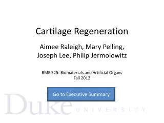
Endogenousstemcells • During embryogenesis most of the renal parenchymal cells arederived from the metanephricmesenchymal cells, which are multipotent and self-renewing. • Invariousanimalmodelsmanyprogenitors of renalcellshavebeenidentified based on their location within the renal compartment includingalong tubular cells, within Bowman's capsule, the papillary region, andwithinthecorticalinterstitium. • In human kidney, theexistence of a population of cells (CD133+/CD24+) located in theurinary pole of the glomerulus.After stimulation they can give rise to tubular epithelialcells as well as podocytes. • Othercellpopulation(CD133+/CD24+/PDX+) present between the urinarypole and the vascular pole that gives rise to podocytes only.
Renal progenitor cells Vascularstalk Detaching podocyte Detaching podocyte Podocyte progenitors Bridge Mature podocytes Renal progenitors Urinary pole
Exogenousstemcells • Stem cells isolated from bonemarrow and amniotic fluid, MSC, have been primarily used asexogenous sources for suitable therapy to treat kidney disease. • Injections of thesestemcellsseemstocause a functionalrecovery of theinjuredkidney. • It is notclearhowthesecellsareabletoactatthe site of degeneratedtissue. • The mechanismscould be transdifferentiation and integrationintotheinjured site. Inthislattercasetheefficiencyvariesbetween 2%-20%. • Inoppositetothislowefficacythetherapy has positiveoutcomes, maybeduetotheautocrine / paracrinemechanismsexertedbysecretedcytokines of theseinjectedcells.
HumoralfactorsreleasedbyprogenitorcellsinvolvedinkidneyrepairHumoralfactorsreleasedbyprogenitorcellsinvolvedinkidneyrepair • Vascularendothelialgrowthfactor (VEGF) attenuatesglomerularinflammation and acceleratesglomerularcapillaryrepair. • Hepatocytegrowthfactor (HGF) is an angiogenicgrowthfactorpreventsepithelialcell death and enhances regeneration and remodeling ofinjured or fibrotic renal tissue. • Insulin-likegrowthfactor (IGF): enhanceglomerularfiltrationrate and mitogenicforproximaltubulecells • Theseeffectswereindividuallyobserved, thejoinedeffectsareunknown.
Microvesicles and cell-cellcommunication • Release of microvesiclesfromcellshavebeenobservedin vivo. • Microvesicles are derived from the endosomalmembrane compartment and after fusion with the plasmamembrane are released from the cell surface. The production of microvesicles is initiatedbycellularactivationbyligands and stresssignals. • Microvesiclescandelivergeneticinformationsuchas mRNA orevenmicroRNA acting as a vehicle for genetic exchangebetweencells. • It is now known thatmicrovesicles may interact with cells through specific receptor–ligandinteractions.
Microvesicles and stemcells • The phenotype of stem cells isreversibly changing during the cell cycle. • The same stem cell may show different phenotypesin different functional states, depending on the cell cyclephase. • This dynamic context is regulated by the microenvironmentand in particular the microvesicle-mediatedtransfer of genetic information between cells. • Microvesiclesgenerated from endothelial progenitor cells (EPCs)when internalized in normal endothelial cells activate anangiogenic program by a horizontaltransfer of mRNA. • The microvesicles released by ESCsare able to reprogram hematopoietic progenitors by deliveryof mRNA and proteins. • Theseindicate that stem cells may release biologically activemicrovesicles not only in their microenvironment butalso in the blood as seen after bone marrow mobilization.
Microvesiclesroleinkidneyrepair • Human MSCsabletoreleasemicrovesiclesand stimulate invitro proliferation and apoptosis resistance of tubular epithelial cells. • Moreover, accelerate in vivo the functional and morphological recovery of tubularcellsin SCID micewithchemicalinduced AKI. • RNasetreatment of microvesicles abolished both the invitro andthe invivo effects of microvesicles, suggesting a mechanismdependenton RNA delivery. • Microvesiclesreleasedfromstemcells at the site of tissue injury may inducededifferentiation of resident cells surviving injury,makingthem transiently acquire a stem cell-like phenotype withthe activation of tissue regenerative programs.
Repair mechanisms of stem cells in kidney regeneration Injured cell-stem cell communication Paracrine soluble mediators EGF, IGF-1, VEGF, MSP, HGF Tubular epitelial cell injury Tissue regeneration Microvesicles with mRNA or miRNA Circulation Differentiation Bone marrow-derived or tissue resident stem cells Stem cell-injured cell communication Paracrine soluble mediators Tubular epithelial cell injury Microvesicles with mRNA or miRNA EGF, IGF-1, VEGF, MSP, HGF De-differentiation Proliferation Re-differentiation Circulation Tubular epithelial cell repair Bone marrow-derived stem cells Tissue resident stem cells
GenetherapyforkidneydiseaseI • Genetherapyforacutekidneyfailure: • During AKF happen a series of eventssuchaslocal inflammation, the expression ofchemotactic and chemoattractant molecules with higher apoptosisrates and impairedregeneration. • BlockingNFkBsignalingpahtwaysinhibitthecytokine/chemokine (MCP1) activationsoabolishtherecruitment of inflammatorycells. • Downregulation of ICAM 1 expressionbyantiseneoligoscandecreasethenumbers of infiltratingneutrophils. • Knock down of complement 3 and upregulation of Bcl2 byadenovirostransfectioncouldrescuerenalcellsfromextendedapoptosis. • Hepatocytegrowthfactor (HGF) therapycanpreventinflammation and organdegeneration.
GenetherapyforkidneydiseaseII • Genetherapyforchronickidneyfailure: • The imbalancebetweentheinflammatory and anti-inflammatoryprocesses is thebasics of chronickidneyfailure. The major player is theTGF-bcytokineinthisinjuryprocesspromotingfibrosiswithcollagendeposition. • Duetoitscomplexsignalling and manytargetsseveralattemptswere made tocontrolTGF-bsignalingespeciallythedownstream SMAD (2/3, 4, 7) molecules. Upregulation of SMAD 7 (bythisinhibiting SMAD2/3 receptors of TGF-b) couldabolishfibrosis and inflammation. • Moreover, targeting AP-1 orNFkBgavealsosuccessfullresults. • Ex vivo genetherapy: • Both regularrenaltransplantation and stemcelltherapycouldhavebenefitfromgenetherapycontrollingimmunogenecity of theallogentissues. Someattemptswere made using CTLA4-Ig and rIL-10 constructs.
Summary • Kidney is theonethe most highlydifferentiatedorganinour body. • Kidneyfailureduetodifferentbackgrounds is increasingin human populaton. • Consideringthesefactsit is importanttoenhancetheconventionalkidneyreplacementsalongwithnoveltherapeuticalapproaches. • Noveldata show thatkidneycan be regeneratedusingartificialscaffoldsalongwithsomaticcells. • Moreover, promisingapplicationsareemergingusingexogenous and endogenousoriginitadstemcellsandvariousgenetherapyforkidneyregeneration.