Cartilage Regeneration
Cartilage Regeneration. BME 525: Biomaterials and Artificial Organs Fall 2012. Aimee Raleigh, Mary Pelling , Joseph Lee, Philip Jermolowitz. Go to Executive Summary. Instructions. This presentation is to be used as an educational tool to learn more about cartilage regeneration

Cartilage Regeneration
E N D
Presentation Transcript
Cartilage Regeneration BME 525: Biomaterials and Artificial Organs Fall 2012 Aimee Raleigh, Mary Pelling, Joseph Lee, Philip Jermolowitz Go to Executive Summary
Instructions • This presentation is to be used as an educational tool to learn more about cartilage regeneration • Hover over words that look like this for additional information. • Pictures with a blue border are links to other sections. • To obtain the best results, follow the instructions here(recommended). Click Here to Continue to the Home Page Home Button References Page Instructions Page Use this to keep track of where you are. Click on greyed sections to revisit. Back Button Title Page > Instructions
Instructions Move your mouse away when you’re done • This presentation is to be used as an educational tool to learn more about cartilage regeneration • Hover over words that look like this for additional information. • Pictures with a blue border are links to other sections. • To obtain the best results, follow the instructions here(recommended). Click Here to Continue to the Home Page Home Button References Page Instructions Page Use this to keep track of where you are. Click on greyed sections to revisit. Back Button Title Page > Instructions
Instructions • This presentation is to be used as an educational tool to learn more about cartilage regeneration • Hover over words that look like this for additional information. • Pictures with a blue border are links to other sections. • To obtain the best results, follow the instructions here(recommended). Move mouse to lower-left corner of the PowerPoint Click on the pen button that appears Go to ‘Arrow Options’ and click ‘Visible’ Click Here to Continue to the Home Page Home Button References Page Instructions Page Use this to keep track of where you are. Click on greyed sections to revisit. Back Button Title Page > Instructions
Cartilage Regeneration: Executive Summary Our presentation will focus on injuries (both acute and chronic) to hyaline cartilage. This cartilage is predominantly found at articulating joint surfaces and serves to lubricate the contacting entities to enable a low friction environment for repetitive articulation of joint motion. Many orthopaedic surgeons have relied on techniques such as microfracture, debridement, and OATSprocedures, which are less than ideal because the regenerated tissue is mechanically inferior to native hyaline cartilage and in most cases leaves the patient in a pre-arthritic state. Recently, there have been huge advances in the development of treatments across a range of backgrounds, including both synthetic biomaterials-based scaffolds and plugs as well as cell-based techniques to mimic the local chondrocyte orientation and structure. While many techniques are used in the clinic today, the highlight of our biomedical vendor profile will be on Carticel, a technique that has been around since 1995. Carticel is a technology based on the autologous biopsy of a patient’s own chondrocytes and the subsequent culture and expansion of these cells for re-implantation during a later surgical procedure. Learn How Use This Module Begin Module Executive Summary
Cartilage Regeneration: Executive Summary Our presentation will focus on injuries (both acute and chronic) to hyaline cartilage. This cartilage is predominantly found at articulating joint surfaces and serves to lubricate the contacting entities to enable a low friction environment for repetitive articulation of joint motion. Many orthopaedic surgeons have relied on techniques such as microfracture, debridement, and OATSprocedures, which are less than ideal because the regenerated tissue is mechanically inferior to native hyaline cartilage and in most cases leaves the patient in a pre-arthritic state. Recently, there have been huge advances in the development of treatments across a range of backgrounds, including both synthetic biomaterials-based scaffolds and plugs as well as cell-based techniques to mimic the local chondrocyte orientation and structure. While many techniques are used in the clinic today, the highlight of our biomedical vendor profile will be on Carticel, a technique that has been around since 1995. Carticel is a technology based on the autologous biopsy of a patient’s own chondrocytes and the subsequent culture and expansion of these cells for re-implantation during a later surgical procedure. A commonly used practice in which small cartilage defects are repaired by drilling tiny holes into the underlying subchondral bone and initiating a blood and bone marrow response. Learn How Use This Module Begin Module Executive Summary
Cartilage Regeneration: Executive Summary Our presentation will focus on injuries (both acute and chronic) to hyaline cartilage. This cartilage is predominantly found at articulating joint surfaces and serves to lubricate the contacting entities to enable a low friction environment for repetitive articulation of joint motion. Many orthopaedic surgeons have relied on techniques such as microfracture, debridement, and OATSprocedures, which are less than ideal because the regenerated tissue is mechanically inferior to native hyaline cartilage and in most cases leaves the patient in a pre-arthritic state. Recently, there have been huge advances in the development of treatments across a range of backgrounds, including both synthetic biomaterials-based scaffolds and plugs as well as cell-based techniques to mimic the local chondrocyte orientation and structure. While many techniques are used in the clinic today, the highlight of our biomedical vendor profile will be on Carticel, a technique that has been around since 1995. Carticel is a technology based on the autologous biopsy of a patient’s own chondrocytes and the subsequent culture and expansion of these cells for re-implantation during a later surgical procedure. An arthroscopic procedure in which the damaged tissue is smoothed and cleansed of debris. This technique is not corrective and is primarily used to delay further injury to the site. Learn How Use This Module Begin Module Executive Summary
Cartilage Regeneration: Executive Summary Our presentation will focus on injuries (both acute and chronic) to hyaline cartilage. This cartilage is predominantly found at articulating joint surfaces and serves to lubricate the contacting entities to enable a low friction environment for repetitive articulation of joint motion. Many orthopaedic surgeons have relied on techniques such as microfracture, debridement, and OATSprocedures, which are less than ideal because the regenerated tissue is mechanically inferior to native hyaline cartilage and in most cases leaves the patient in a pre-arthritic state. Recently, there have been huge advances in the development of treatments across a range of backgrounds, including both synthetic biomaterials-based scaffolds and plugs as well as cell-based techniques to mimic the local chondrocyte orientation and structure. While many techniques are used in the clinic today, the highlight of our biomedical vendor profile will be on Carticel, a technique that has been around since 1995. Carticel is a technology based on the autologous biopsy of a patient’s own chondrocytes and the subsequent culture and expansion of these cells for re-implantation during a later surgical procedure. (OsteochondralAutograft Transfer System) Involves the coring out of a patient’s own articular cartilage from a nonweight-bearing site and inserting these native cartilage plugs into the tissue defect Learn How Use This Module Begin Module Executive Summary
Cartilage Regeneration: Executive Summary Our presentation will focus on injuries (both acute and chronic) to hyaline cartilage. This cartilage is predominantly found at articulating joint surfaces and serves to lubricate the contacting entities to enable a low friction environment for repetitive articulation of joint motion. Many orthopaedic surgeons have relied on techniques such as microfracture, debridement, and OATSprocedures, which are less than ideal because the regenerated tissue is mechanically inferior to native hyaline cartilage and in most cases leaves the patient in a pre-arthritic state. Recently, there have been huge advances in the development of treatments across a range of backgrounds, including both synthetic biomaterials-based scaffolds and plugs as well as cell-based techniques to mimic the local chondrocyte orientation and structure. While many techniques are used in the clinic today, the highlight of our biomedical vendor profile will be on Carticel, a technique that has been around since 1995. Carticel is a technology based on the autologous biopsy of a patient’s own chondrocytes and the subsequent culture and expansion of these cells for re-implantation during a later surgical procedure. Cartilage cell Learn How Use This Module Begin Module Executive Summary
Cartilage Regeneration: Executive Summary Our presentation will focus on injuries (both acute and chronic) to hyaline cartilage. This cartilage is predominantly found at articulating joint surfaces and serves to lubricate the contacting entities to enable a low friction environment for repetitive articulation of joint motion. Many orthopaedic surgeons have relied on techniques such as microfracture, debridement, and OATSprocedures, which are less than ideal because the regenerated tissue is mechanically inferior to native hyaline cartilage and in most cases leaves the patient in a pre-arthritic state. Recently, there have been huge advances in the development of treatments across a range of backgrounds, including both synthetic biomaterials-based scaffolds and plugs as well as cell-based techniques to mimic the local chondrocyte orientation and structure. While many techniques are used in the clinic today, the highlight of our biomedical vendor profile will be on Carticel, a technique that has been around since 1995. Carticel is a technology based on the autologous biopsy of a patient’s own chondrocytes and the subsequent culture and expansion of these cells for re-implantation during a later surgical procedure. Derived or transferred from the same individual Learn How Use This Module Begin Module Executive Summary
Main Menu – Cartilage Regeneration Executive Summary Cartilage Overview Disease States Commercial and Research Techniques Material Selection and Design Implant Performance and Failure Modes Acknowledgements and References Future Directions Title Page > Instructions > Main Menu
Main Menu – Cartilage Regeneration Executive Summary Cartilage Overview Disease States Commercial and Research Techniques Material Selection and Design Implant Performance and Failure Modes Acknowledgements and References Future Directions Title Page > Instructions > Main Menu
Main Menu – Cartilage Regeneration Executive Summary Cartilage Overview Disease States Commercial and Research Techniques Material Selection and Design 1 Implant Performance and Failure Modes Acknowledgements and References Future Directions Title Page > Instructions > Main Menu
Main Menu – Cartilage Regeneration Executive Summary Cartilage Overview Disease States Commercial and Research Techniques Material Selection and Design 2 Implant Performance and Failure Modes Acknowledgements and References Future Directions Title Page > Instructions > Main Menu
Main Menu – Cartilage Regeneration Executive Summary Cartilage Overview Disease States Commercial and Research Techniques Material Selection and Design 3 Implant Performance and Failure Modes Acknowledgements and References Future Directions Title Page > Instructions > Main Menu
Main Menu – Cartilage Regeneration Executive Summary Cartilage Overview Disease States Commercial and Research Techniques Material Selection and Design 4 Implant Performance and Failure Modes Acknowledgements and References Future Directions Title Page > Instructions > Main Menu
Main Menu – Cartilage Regeneration Executive Summary Cartilage Overview Disease States Commercial and Research Techniques Material Selection and Design 5 Implant Performance and Failure Modes Acknowledgements and References Future Directions Title Page > Instructions > Main Menu
Main Menu – Cartilage Regeneration Executive Summary Cartilage Overview Disease States Commercial and Research Techniques Material Selection and Design 6 Implant Performance and Failure Modes Acknowledgements and References Future Directions Title Page > Instructions > Main Menu
Cartilage: 3 Main Types Intervertebral Disc: White Fibrocartilage Outer Ear, Larynx, and Epiglottis: Elastic Cartilage Articulating Joint Surfaces: Hyaline Cartilage 9 7 10 Continue to Background 8 Background
Background-Question1 Elastic cartilage’s best property is? • It heals very rapidly • It’s transparent • Its ability to stretch and then return to its normal shape • It’s incredibly tough and resistant to impact Background > Quiz
Background-Question1 Elastic cartilage’s best property is? • It heals very rapidly • It’s transparent • Its ability to stretch and then return to its normal shape • It’s incredibly tough and resistant to impact Go to Question 2 Background > Quiz
Background-Question1 Elastic cartilage’s best property is? • It heals very rapidly • It’s transparent • Its ability to stretch and then return to its normal shape • It’s incredibly tough and resistant to impact Go to Question 2 Background > Quiz
Background-Question1 Elastic cartilage’s best property is? • It heals very rapidly • It’s transparent • Its ability to stretch and then return to its normal shape • It’s incredibly tough and resistant to impact Go to Question 2 Background > Quiz
Background-Question1 Elastic cartilage’s best property is? • It heals very rapidly • It’s transparent • Its ability to stretch and then return to its normal shape • It’s incredibly tough and resistant to impact Go to Question 2 Background > Quiz
Background-Question 2 Which of the following body parts would most likely have elastic cartilage? • Intervertebral discs • Ear • Fingernails • Joints Background > Quiz
Background-Question 2 Which of the following body parts would most likely have elastic cartilage? • Intervertebral discs • Ear • Fingernails • Joints Go to Question 3 Background > Quiz
Background-Question 2 Which of the following body parts would most likely have elastic cartilage? • Intervertebral discs • Ear • Fingernails • Joints Go to Question 3 Background > Quiz
Background-Question 2 Which of the following body parts would most likely have elastic cartilage? • Intervertebral discs • Ear • Fingernails • Joints Go to Question 3 Background > Quiz
Background-Question 2 Which of the following body parts would most likely have elastic cartilage? • Intervertebral discs • Ear • Fingernails • Joints Go to Question 3 Background > Quiz
Background-Question 3 What are the only kinds of cells found in cartilage? • Chondrocytes • Monocytes • Phagocytes • Hemocytes Background > Quiz
Background-Question 3 What are the only kinds of cells found in cartilage? • Chondrocytes • Monocytes • Phagocytes • Hemocytes Go to Question 4 Background > Quiz
Background-Question 3 What are the only kinds of cells found in cartilage? • Chondrocytes • Monocytes • Phagocytes • Hemocytes Go to Question 4 Background > Quiz
Background-Question 3 What are the only kinds of cells found in cartilage? • Chondrocytes • Monocytes • Phagocytes • Hemocytes Go to Question 4 Background > Quiz
Background-Question 3 What are the only kinds of cells found in cartilage? • Chondrocytes • Monocytes • Phagocytes • Hemocytes Go to Question 4 Background > Quiz
Background-Question 4 What is the primary function of fibrocartilage? • To act as a wear surface • To reflect sound • To absorb nutrients • To absorb shock Background > Quiz
Background-Question 4 What is the primary function of fibrocartilage? • To act as a wear surface • To reflect sound • To absorb nutrients • To absorb shock Go to Question 5 Background > Quiz
Background-Question 4 What is the primary function of fibrocartilage? • To act as a wear surface • To reflect sound • To absorb nutrients • To absorb shock Go to Question 5 Background > Quiz
Background-Question 4 What is the primary function of fibrocartilage? • To act as a wear surface • To reflect sound • To absorb nutrients • To absorb shock Go to Question 5 Background > Quiz
Background-Question 4 What is the primary function of fibrocartilage? • To act as a wear surface • To reflect sound • To absorb nutrients • To absorb shock Go to Question 5 Background > Quiz
Background-Question 5 Which of the following materials properties can be most closely associated with hyaline cartilage? • It experiences creep • It’s anisotropic • It’s electrically conductive • It has a high coefficient of expansion Background > Quiz
Background-Question 5 Which of the following materials properties can be most closely associated with hyaline cartilage? • It experiences creep • It’s anisotropic • It’s electrically conductive • It has a high coefficient of expansion Return to Background Background > Quiz
Background-Question 5 Which of the following materials properties can be most closely associated with hyaline cartilage? • It experiences creep • It’s anisotropic • It’s electrically conductive • It has a high coefficient of expansion Return to Background Background > Quiz
Background-Question 5 Which of the following materials properties can be most closely associated with hyaline cartilage? • It experiences creep • It’s anisotropic • It’s electrically conductive • It has a high coefficient of expansion Return to Background Background > Quiz
Background-Question 5 Which of the following materials properties can be most closely associated with hyaline cartilage? • It experiences creep • It’s anisotropic • It’s electrically conductive • It has a high coefficient of expansion Return to Background Background > Quiz
3 Main Types 11 12 13 Fibrocartilage Hyaline Cartilage Elastic Cartilage Return to Main Menu Take Background Quiz Background > Selection
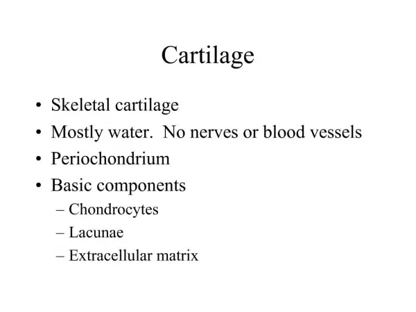
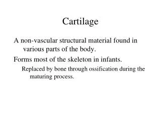
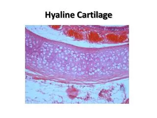
Articular Hyaline Cartilage Hyaline Cartilage is a dense, thin (1-6 mm) connective tissue that is translucent and white and found on most joint surfaces in the body, on the ventral side of ribs, and in the larynx, trachea, and bronchi. The tissue is relatively isolated and lacks blood vessels, lymphatic channels, and neurological innervation. Thus, once this tissue becomes injured, it is difficult for the body to promote an adequate healing response. 14 For our presentation, we will focus on hyaline cartilage, since its presence at articulating joint surfaces makes it especially prone to degradation and thus is an ideal candidate for a biomaterials solution to wear and tear. Molecular Basis Mechanics Return to Overview Cartilage Slide Background > Selection > Hyaline
Molecular Basis of Hyaline Cartilage Hyaline cartilage is a biphasic material that consists of an interstitial fluid phase (mostly water) in combination with an extracellular organic matrix composed of collagen (type II) fibers and proteoglycans. Tissue Ingredient #1: Collagen The arrangement of collagen starts at the nanoscale with procollagen polypeptide chains that form the tropocollagen, and builds up to bundles in the following order: TropocollagenFibrilsFine Collagen FibersFibersBundles This structural redundancy of collagen forms lends to a high tensile strength that endows the matrix of cartilage to adjust to different types of loading conditions. While the collagen in articular collagen is metabolically inert, PGs actually have a relatively fast turnover rate. Thus, collagen is more susceptible to fatigue failure. It undergoes a whole lifetime of compressive cycles and motions, while many components of the extracellular matrix do not. 15 The innate structural complexity of cartilage components makes it an extremely difficult material to replicate with biomaterials! Go to Molecular Basis II Background > Selection > Hyaline > Histology
Molecular Basis of Hyaline Cartilage Hyaline cartilage is a biphasic material that consists of an interstitial fluid phase (mostly water) in combination with an extracellular organic matrix composed of collagen (type II) fibers and proteoglycans. Tissue Ingredient #1: Collagen The arrangement of collagen starts at the nanoscale with procollagen polypeptide chains that form the tropocollagen, and builds up to bundles in the following order: TropocollagenFibrilsFine Collagen FibersFibersBundles This structural redundancy of collagen forms lends to a high tensile strength that endows the matrix of cartilage to adjust to different types of loading conditions. While the collagen in articular collagen is metabolically inert, PGs actually have a relatively fast turnover rate. Thus, collagen is more susceptible to fatigue failure. It undergoes a whole lifetime of compressive cycles and motions, while many components of the extracellular matrix do not. 15 The innate structural complexity of cartilage components makes it an extremely difficult material to replicate with biomaterials! Go to Molecular Basis II Cells do not turnover Background > Selection > Hyaline > Histology
Molecular Basis of Hyaline Cartilage II Tissue Ingredient #2: Proteoglycans PGs consist of large protein-polysaccharide moieties containing a protein core to which various glycosaminoglycans attach in a “bottle brush” arrangement. The collagen retains the PGs, and the PGs retain the water. The collagen fibers limit the PGs to imbibe water to control excess swelling. Under compressive loads, the PG gel attempts to translate to an area of lower pressure, but is restricted by the collagen structure. Therefore, collagen fibers also experience tensile stress when the cartilage is loaded. Keratin Sulfate and Chondroitin Sulfate (two different types of glycosaminoglycans) contain a high concentration of stationary negative chargesthroughout their molecules. These forces imbue the collagen-PG network with rigidity and compressive strength that complements the high tensile strength of the collagen fibers. Go to Molecular Basis III 16 Background > Selection > Hyaline > Histology 2
Molecular Basis of Hyaline Cartilage II The aggregation of these proteoglycan units adds structural support and stiffness to the collagen matrix. Tissue Ingredient #2: Proteoglycans PGs consist of large protein-polysaccharide moieties containing a protein core to which various glycosaminoglycans attach in a “bottle brush” arrangement. The collagen retains the PGs, and the PGs retain the water. The collagen fibers limit the PGs to imbibe water to control excess swelling. Under compressive loads, the PG gel attempts to translate to an area of lower pressure, but is restricted by the collagen structure. Therefore, collagen fibers also experience tensile stress when the cartilage is loaded. Keratin Sulfate and Chondroitin Sulfate (two different types of glycosaminoglycans) contain a high concentration of stationary negative chargesthroughout their molecules. These forces imbue the collagen-PG network with rigidity and compressive strength that complements the high tensile strength of the collagen fibers. Go to Molecular Basis III 16 Background > Selection > Hyaline > Histology 2