CARTILAGE
CARTILAGE . Dr Iram Tassaduq. CARTILAGE . Cartilage is a type of connective tissue derived from mesenchyme, in which the ground substance is hardened and provides support. . CARTILAGE .


CARTILAGE
E N D
Presentation Transcript
CARTILAGE Dr Iram Tassaduq
CARTILAGE • Cartilage is a type of connective tissue derived from mesenchyme, in which the ground substance is hardened and provides support.
CARTILAGE • It is a form of connective tissue composed of cells called chondrocytes and a highly specialized extracellular matrix
CARTILAGE • Matrix is solid and firm • Does not contain vessels or nerves. • Is surrounded by a layer of dense connective tissue, the perichondrium
FUNCTIONS OF CARTILAGE TISSUE • Firm consistency of the extracellular matrix allows the tissue to bear mechanical stresses without permanent distortion • Supports soft tissues. • Shock-absorbing because it is resilient. • Smooth surface allows sliding . • Essential for growth, development of bone.
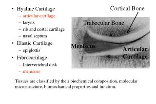
TYPES OF CARTILAGE • Hyaline cartilage • Elastic cartilage • Fibrous cartilage
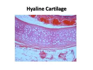
HYALINE CARTILAGE • Cells embedded in clear matrix(hyalos meaning glass) • Perichondrium on surface • Chondrocytes are located in lacunae
HYALINE CARTILAGE • Bluish white color. • Strong, rubbery, flexible tissue.
CHONDROCYTES • Produce and maintain extra cellular matrix. • Either single or in isogenous groups . • Fat droplets, glycogen granules are found in cytoplasm. • Active ones are more basophilic.
TERRITORIAL MATRIX • Contain high concentration of bound sulfate so stains with basic dyes. • Rich in water(70-80%) • Three classes of molecules are present; Collagen, Proteoglycans and glycoproteins.
INTERTERRITORIAL MATRIX • Region that surrounds the territorial matrix • Sulphated proteoglycans are in low concentration
LOCATION Found at • ends of bones • nose • trachea • larynx
PERICHONDRIUM • Dense CT that covers cartilage • Contains blood, nerve supply, lymphatics. • Source of new cartilage cells
PERICHONDRIUM • Divided into two layers Inner cellular Outer fibrous
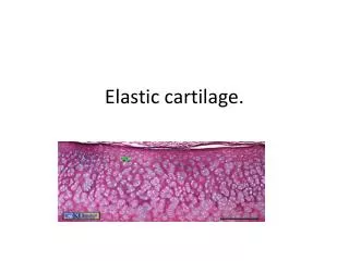
ELASTIC CARTILAGE • Similar to hyaline cartilage but has elastic fibers running in all directions in addition to collagen.
ELASTIC CARTILAGE • The properties of elastin and the fibers it forms give elastic cartilage its ability to be deformed and to spring back into shape immediately. The chondrocytes are more tightly packed together than is the case in hyaline cartilage
ELASTIC CARTILAGE • Elastic cartilage is found in • pinna of the Ear • walls of the Eustachian tube • Epiglottis all places in which the maintenance of a specific shape is important to proper function
FIBROCARTILAGE • Chondrocytes with dense connective tissue. • No surrounding perichondrium. • Rounded nuclei belong to chondrocytes. • In fibrous areas some nuclei are flat, they are of fibroblasts. • Contain type I and type II collagen.
FIBROCARTILAGE • Chondrocytes may lie singly or in pairs, but most often they form short rows between dense bundles of collagen fibers.
FIBROCARTILAGE • Collagen type I is dominant in fibrous cartilage
FIBROCARTILAGE is typically found in relation to joints • intra-articular lips, disks and menisci • intervertebral disks • symphysis pubis.
GROWTH OF CARTILAGE • Growth is attributable to two processes: • Interstitial growth • Mitotic division of preexisting chrondrocytes • Synthesis of matrix • Expands cartilage matrix from within • Occurs in epiphyseal plates, articular cartilage • Appositional growth • Differentiation of perichondrial cells chondroblasts • Synthesis of matrix • Increase in girth
REPAIR OF HYALINE CARTILAGE • Can tolerate considerable amount of stress • Limited ability to repair Because of 1. Immobility of chodrocytes 2. Less ability of chodrocytes to proliferate 3. Avascularity 4. When hyaline cartilage calcifies it is replaced by bone