Download
1 / 29
300 likes | 1.37k Vues
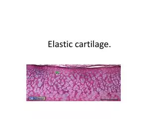
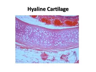
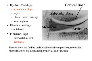
Cortical Bone. Trabecular Bone. Hyaline Cartilage articular cartilage larynx rib and costal cartilage nasal septum Elastic Cartilage epiglottis Fibrocartilage Intervertebral disk meniscus. Meniscus. Articular Cartilage.

E N D
Cortical Bone Trabecular Bone • Hyaline Cartilage • articular cartilage • larynx • rib and costal cartilage • nasal septum • Elastic Cartilage • epiglottis • Fibrocartilage • Intervertebral disk • meniscus Meniscus Articular Cartilage Tissues are classified by their biochemical composition, molecular microstructure, biomechanical properties and function.
AC/Meniscus Functions: • Support large loads • gymnastics • Walking • Lubrication Interested in these structures because when they “breakdown” we get osteoarthritis
Articular Cartilage • Important to understand • Mechanical properties of normal cartilage • Manner by which biochemical and structural factors contribute to the material properties of cartilage • Manner by which changes in tissue composition affect the mechanical properties of cartilage
Diarthrodial joint • Fibrous capsule • Inside lined with synovium which secretes synovial fluid
Microstructure (Solid and Fluid Phase) • Interstitial water • Articular cartilage 68-85%, meniscus 60-70%
Interstitial Water • Constant with age • Increases with OA or degeneration • Amount of water is dependent on
Interstitial water • Ions- • As tissue is compressed-Frictional drag force on walls of the pores of the solid matrix due to interstitial fluid flow through the pores of collagen-PG matrix
Microstructure (Solid and Fluid Phase) • Collagen • Proteoglycans • Cells No blood or nerves in cartilage
Collagen: made up of molecules (tropocollagen--1.4 nm) that polymerize to form fibrils • Type II (AC), forms bundles, with diam.=2 to 10 microns • Type I (meniscus), forms fibrils, with diam. = 20-200 nm
Proteoglycan: protein with bound side chains (glycosaminoglycans)
Proteoglycans • Negative charge attracts +ions (K and Na) • Swelling pressure • PG want to be 5-10 times larger, but not enough room in cartilage
Material Properties • Steel is linear elastic (E,) • Soft tissues ARE NOT!! • Water movement (forces depend on rate-damping) STEEL
Material Properties • Viscoelastic behavior are dominated by frictional drag of interstitial fluid flow through the porous collagen-proteoglycan solid matrix, thus causing viscous dissipation
Material Properties-Anisotropy/Inhomogeneous • Transversely Isotropic • Inhomogeneous
Constitutive Equation: • Linear Elastic Materials (Steel) • Hookes’ Law: = E • Viscoelastic materials (AC/meniscus) • Biphasic Theory (2 phase) • Triphasic Theory (3 phase)
Deform. Force time time Tensile Stress Relaxation Test Tension • Equilibrium Tensile Modulus (1-30 MPa) • Type of tissue • Age of animal • Type of joint • Sample location • Depth of sample (surface = 10MPa, Middle =4.5MPa) • Relative orientation • Biochemical comp/ molecular structure • State of degeneration (Normal =10MPa, OA=1.4MPa)
Compression • Compressive Aggregate Modulus (HA)(0.4-1.5 MPa) Force Deform time time Confined Compression Creep Test
Compression • HA varies inversely with water content *OA patients have increased water • HA varies directly with PG content • Not dependent on collagen content
Methods of Failure-Cartilage Fracture – Fracture with Bone Wear Degeneration Blunt Trauma (intense compression and shear forces) Bone Bone
Methods of Failure - Meniscus • Degeneration • Tearing