Understanding Cartilage: Composition, Function, and Growth Mechanisms
Cartilage is a specialized form of connective tissue found in various parts of the body, providing support and facilitating bone movement. Learn about its composition, characteristics, growth methods, and functions. Explore the types of cartilage, such as hyaline, elastic, and fibrocartilage, along with their examples and functions.

Understanding Cartilage: Composition, Function, and Growth Mechanisms
E N D
Presentation Transcript
Cartilage Dr.PARDEEP KUMAR
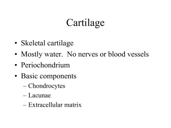
CARTILAGE • It is a specialized form of connective tissue consists of cells, fibres & ground substance. The cells are embedded in the intercellular matrix • Origin - Mesenchymal in origin.
Cartilage • Embryo • More prevalent than in adult • Skeleton initially mostly cartilage • Bone replaces cartilage in fetal and childhood periods
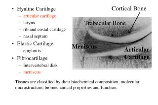
Location of cartilage in adults • External ear • Nose • “Articular” – covering the ends of most bones and movable joints • “Costal” – connecting ribs to sternum • Larynx - voice box
Epiglottis – flap keeping food out of lungs • Cartilaginous rings holding open the air tubes of the respiratory system (trachea and bronchi) • Intervertebral discs • Pubic symphysis • Articular discs such as meniscus in knee joint
Composition: • 1) Cells: Chondrocytes, Chondroblasts • 2) Fibres: White fibers and yellow elastic fibres. • 3) Ground substance: Consists of Glyco-samino-glycans: • i) Chondroitin sulfate. • ii) Kerato sulfate. • iii) Hyaluronic acid.
Characteristics of cartilage • Avascular, devoid of lymphatics & nerves. Gets its nutrition from blood vessels of adjacent tissue by process of diffusion • Its matrix is not calcified ( Cartilage turn into bone by calcification) • Cartilage grows by 2 methods • Interstitial (endogenous method) • Appositional (exogenous growth) • It forms skeleton of body in early life, most of which is replaced by bone in adult
Growth of a cartilage Cartilage grows by 2 methods • Interstitial growth: Expansion of internal mass of cartilage by division of chondrocyte ("in the middle“) Increasing in LENGTH; chondrocytes divide and secrete matrix from w/in lacunae • Appositional growth: when cartilage grows by adding new layer on its surface by the cells of perichondium ("at the edge“) • Increasing in WIDTH; chondroblasts deposit matrix on surface of pre-existing cartilage
Functions of Cartilage • Supports soft tissues of body • Facilitates bone movement by virtue of its smooth surface Nutrition of Cartilage • Cartilage is avascular • Hyaline & elastic cartilage gets nutrition from vessels of Perichondrium • Fibro cartilage gets nutrition from blood vessels of surrounding connective tissue & articular cartilage from synovial fluid
Perichondrium ( covering of cartilage) • Dense fibrous connective tissue which encloses cartilage is called Perichondrium • It is essential for the growth & maintenance of cartilage
CHONDROCYTES & CHONDROBLASTS • They are cartilage cells, irregular in shape. • The almond shaped spaces they occupy are lacunae. • They may remain in ground substance widely separated as discrete or in groups. • They are developed from mesenchymal cells.
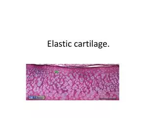
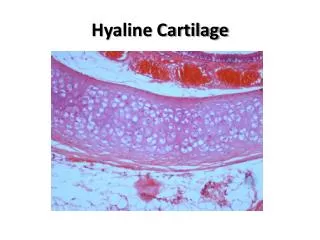
Types of cartilage: 3 • Hyaline cartilage: flexible and resilient • Chondrocytes appear spherical • Lacuna – cavity in matrix holding chondrocyte • Collagen the only fiber • Elastic cartilage: highly bendable • Matrix with elastic as well as collagen fibers • Epiglottis, larynx and outer ear • Fibrocartilage: resists compression and tension • Rows of thick collagen fibers alternating with rows of chondrocytes (in matrix) • Knee menisci and annunulus fibrosis of intervertebral discs
Types: Three types of CARTILAGE (1) Hyaline cartilage: • It is translucent so called hyaline • Fibres are scanty, relatively cellular i,e more Chondrocytes with more ground substance. • FUNCTION • Support tissue and organs
Examples of Hyaline Cartilage: • Articular cartilage • Costal cartilage • Trachea, bronchi, • Nasal cartilage etc
(2) White fibro-cartilage: • It contains dense white fibrous tissueChondrocytes in more collagen fibre, few ground substance. • FUNCTION • Support with great tensile strength
(2) White fibro-cartilage: Examples: • Inter vertebral disc, • Glenoid & Acetabular Labrum • Pubic symphysis etc
Intervertebral Discs • It is a fibrocartilaginous disc, situated between the bodies of 2 vertebrae & held to them by means of ligament.
It has 2 component Annulus fibrous / outer cartilaginous • It has external layer of dense connective tissue, but is mainly composed of overlapping lamina of cartilage Nucleus pulposus/ Inner liquid • It is situated in the centre of annulus fibrosus. It consists of few rounded cells embedded in amorphous fluid rich in hyaluronic acid
Functions • They allow certain amount of movement between vertebrae • They act as shock absorber • They prevent friction between corresponding vertebrae • They contribute about 1/5th of total length of the vertebral column
(3) Yellow elastic cartilage: • It is a modified elastic connective tissue in which elastic fibers are numerous • More yellow elastic fibres, Chondrocytes in scanty ground substance. Examples: • External ear, • Epiglottis, • Auditory tube, • Some laryngeal cartilage • FUNCTION • Support with flexibility