Download
1 / 13
160 likes | 838 Vues
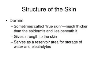
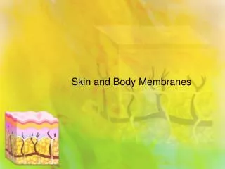
Chapter 4 Skin and Body Membranes “The Dermis”. Dermis. Two layers 1. Papillary layer Projections called dermal papillae Pain receptors Capillary loops 2. Reticular layer Blood vessels Glands Nerve receptors. Skin Structure. Normal Skin Color Determinants. Melanin

E N D
Dermis Two layers 1. Papillary layer • Projections called dermal papillae • Pain receptors • Capillary loops 2. Reticular layer • Blood vessels • Glands • Nerve receptors
Normal Skin Color Determinants Melanin • Yellow, brown or black pigments Carotene • Orange-yellow pigment from some vegetables Hemoglobin • Red coloring from blood cells in dermis capillaries • Oxygen content determines the extent of red coloring
Appendages of the Skin Sebaceous glands • Produce oil - Lubricant for skin - Kills bacteria • Most with ducts that empty into hair follicles • Glands are activated at puberty
Free nerve endings detect temp, mechanical stimuli (such as pressure), pain, & touch Meissner’s corpuscles Tactile, light touch, found in abundance in fingertips, palms, soles, lips, tongue, face, genitals. In dermal papillae. Pacinian corpuscles Deep, pressure, adaptive Nerve Receptors
Appendages of the Skin Sweat glands • Widely distributed in skin • Two types 1. Eccrine • Open via duct to pore on skin surface 2. Apocrine • Ducts empty into hair follicles
Sweat and Its Function Composition • Mostly water • Some metabolic waste • Fatty acids and proteins (apocrine only) Function • Helps dissipate excess heat • Excretes waste products • Acidic nature inhibits bacteria growth Odor is from associated bacteria
Appendages of the Skin Hair • Produced by hair bulb • Consists of hard keratinized epithelial cells • Melanocytes provide pigment for hair color Figure 4.7c
Hair Anatomy • Central medulla • Cortex surrounds medulla • Cuticle on outside of cortex -Most heavily keratinized
Associated Hair Structures Hair follicle • Dermal and epidermal sheath surround hair root Arrector pilli • Smooth muscle • Goose bumps Sebaceous gland Sweat gland
Appendages of the Skin Nails • Scale-like modifications of the epidermis - Heavily keratinized • Stratum basale extends beneath the nail bed - Responsible for growth • Lack of pigment makes them colorless
Nail Structures • Free edge • Body • Root of nail • Eponychium – proximal nail fold that projects onto the nail body